


CT may be superior to MRI for pituitary tumor detection
1. Among patients with surgically-confirmed functional pituitary microadenomas, dynamic contrast-enhanced multisection computed tomography (DCE-MCT) was associated with improved tumor detection when compared with magnetic resonance (MR) imaging.
Evidence Rating: 3 (Average)
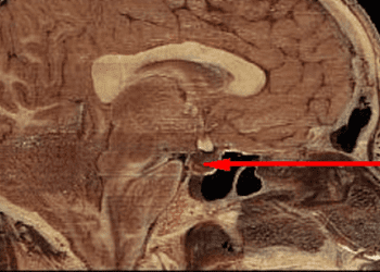
Study Rundown: Pituitary microadenomas are small tumors, less than 10mm in diameter, that can be difficult to localize without the aid of imaging. The current diagnostic standard-of-care is MR imaging, but this technique often lacks the temporal resolution to allow for surgical planning prior to resection. DCE-MCT—a separate technique incorporating information about imaging changes over time—can help provide the spatial and temporal resolution needed to better visualize these small lesions, but it has not been formally validated for this use. In this retrospective cohort study, a small group of patients with surgically-confirmed functional pituitary microadenomas underwent both MR imaging and DCE-MCT to delineate the effectiveness of DCE-MCT in aiding with the detection of pituitary microadenomas. DCE-MCT was able to successfully identify the target lesion in nearly all cases, whereas MR imaging failed in nearly half of the cases. While this study was not a direct comparison of the two techniques , it suggests that there are important differences between MR and CT imaging with regard to pituitary microadenoma evaluation. This study was limited by its overall small sample size and by the lack of standardization of the MR imaging modalities used. Future studies would benefit from a larger, head-to-head comparison of dynamic contrast-enhanced MR and CT imaging in the evaluation of small pituitary tumors.
Click to read the study in American Journal of Neuroradiology
In-Depth [retrospective cohort]: This study evaluated consecutive cases of functional pituitary microadenomas over a 10-year period from 2004-2010. Preoperative MR imaging and DCE-MCT was done on all patients, and tumor size and location was confirmed by trans-sphenoidal endoscopic surgery in all cases. Image reconstruction of the contrast agent dynamics was done using a Matlab-based analysis to evaluate the voxelwise differences between normal pituitary gland and pituitary microadenoma. The overall sensitivity for tumor detection was 92.9% (26/28) with DCE-MCT as compared to 53.8% (15/28) with MR imaging. Microadenomas secreting adrenocorticotropic hormone (ACTH), growth hormone, and prolactin were all identified, with the greatest relative improvement in prediction using DCE-MCT found in ACTH microadenomas, correctly identifying 11 of 13 cases as opposed to only 4 of 13 using MR imaging.
More from this author: Intraarterial therapy improves outcomes in acute ischemic stroke, Meta-analysis suggests endovascular therapy improves stroke outcomes, Magnetic resonance imaging may aid detection of pulmonary hypertension, Breast ultrasound may improve cancer detection in dense breasts, Optical molecular imaging improves lesion localization during liver procedures
Image: CC/davenyin/Wikimedia
©2014 2 Minute Medicine, Inc. All rights reserved. No works may be reproduced without expressed written consent from 2 Minute Medicine, Inc. No article should be construed as medical advice and is not intended as such by the authors, editors, staff or by 2 Minute Medicine, Inc.
RelatedReports