


Epileptogenic foci may be lateralized using functional brain glutamate imaging
1. Among patients with drug-resistant temporal lobe epilepsy not associated with a brain lesion, focally elevated levels of glutamate were detected to correctly lateralize the epileptogenic focus using glutamate chemical exchange saturation transfer (GluCEST) imaging.
2. No significant differences in hippocampal volumes were detected between the hemispheres of affected patients, and among control patients, glutamate levels were not significantly different between the left and right hippocampi.
Evidence Rating Level: 3 (Fair)
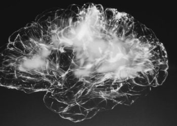
Study Rundown: Epilepsy encompasses a broad spectrum of neurological disturbances symptomatically marked by recurrent seizures. Seizures characteristically occur secondary to focal or generalized neuronal hyperexcitation, typically mediated by glutamate, the major excitatory neurotransmitter. While the true cause of the seizure may be a loss of neuronal inhibition, typically mediated by gamma aminobutyric acid (GABA), the net result may often be consistently elevated levels of glutamate at the epileptogenic focus. Elevated levels of glutamate have in fact been reported in animal models of epilepsy, revealing increased extracellular glutamate buildup associated with epileptogenesis secondary to neuronal hyperexcitation, and may even be mechanistically related to the long-term sequelae of repeated seizures by way of excitotoxicity, or neural loss secondary to excess stimulation. Despite extensive research into the mechanisms underlying epilepsy and the resultant pharmacotherapeutic innovations, approximately one in three patients with epilepsy do not respond to medication alone and may subsequently require MRI-guided surgical removal of the seizure-generating section of the temporal lobe or hippocampus. However, a significant portion of drug-resistant patients will show no obvious causative lesion. The present study examined these patients with drug-resistant, nonlesional epilepsy using a novel magnetic resonance imaging (MRI) technique, glutamate chemical exchange saturation transfer (GluCEST), to characterize glutamate concentrations throughout the brain as a biomarker for anatomical localization of the epileptogenic focus. Using GluCEST imaging, hippocampal glutamate levels were found to be elevated ipsilateral to the epileptogenic hemisphere in all subjects as confirmed by intracranial EEG, and in one patient, postoperative results and pathology. Differences in glutamate concentrations between the hemispheres was not seen in control subjects, and findings regarding glutamate concentrations in both control and epileptic subjects were confirmed by MR spectroscopy. This study introduced a novel glutamate imaging technique which may be of value not just in seizure localization for a group of patients previously lacking anatomic information to guide therapy, but also for potential use in diseases marked by perturbations in gross glutamate concentration within the brain. However, conclusions from this study are limited by its small sample size of four patients and lack of broader post-surgical confirmation, necessitating a significantly larger study, though further data collection was ongoing at the time of publication.
Click to read the study in Science Translational Medicine
Relevant Reading: Gene expression of glutamate metabolizing enzymes in the hippocampal formation in human temporal lobe epilepsy
In-Depth [prospective cohort]: The researchers in this study developed the 7T-MRI based GluCEST imaging method to measure glutamate and used this technique to assess its levels in the hippocampus. Activity within these bilateral structures nested within the temporal lobes is frequently associated with seizure onset, most typically in mesial temporal sclerosis, which may rarely be difficult to visualize on structural MRI. GluCEST imaging with a 7-T MRI scanner was performed in four medication-resistant patients (1 male, age 47; 3 female, mean age: 40 years) with MRI-confirmed nonlesional temporal lobe epilepsy and 11 control patients (3 male and 8 female; mean age: 35 years) without seizure disorders. Each patient in the medication-resistant group had tried and failed between two and seven antiepileptic medications; exclusion criteria included identifiable epilepsy risk factors including head trauma, past history of febrile seizures and family history of seizure. Imaging results were independently analyzed by two epileptologists who were blinded to patient information. Both raters correctly identified hemisphere of seizure onset and interpreted that glutamate levels as detected by GluCEST were higher in the ipsilateral hippocampus than in the contralateral hippocampus. Paired t test for means on the control (two-tailed) and one-tailed t test for epilepsy subjects and indicated higher hippocampal glutamate concentrations at the epileptogenic site (one-tail, P = 0.011), and more specifically within the CA1 hippocampal head region (one-tail, P = 0.03). These findings were verified via intracranial electroencephalography and magnetic resonance spectra. Asymmetric glutamate localization to the hemisphere of epileptic activity was not found in the control group, which showed no significant differences in glutamate across two hemispheres. Interestingly, in one of the medication-resistant subjects, asymmetric lateralization of glutamate level was detected in the hippocampus using magnetic resonance spectroscopy as well, lending additional support for the role of elevated glutamate in seizure pathology. A single patient underwent right temporal lobectomy after intracranial EEG monitoring confirmed the epileptogenic focus revealed by GluCEST imaging, and subsequent pathology revealed findings consistent with mesial temporal sclerosis despite that no lesion was visible on anatomic neuroimaging.
Image: PD
©2015 2 Minute Medicine, Inc. All rights reserved. No works may be reproduced without expressed written consent from 2 Minute Medicine, Inc. Inquire about licensing here. No article should be construed as medical advice and is not intended as such by the authors or by 2 Minute Medicine, Inc.
RelatedReports
