
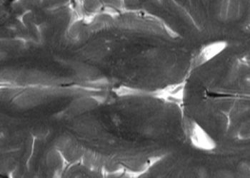
MRI superior to CT for liver cancer diagnosis
1. In patients with chronic liver disease, magnetic resonance (MR) imaging was more sensitive than computed tomography (CT) for the diagnosis of hepatocellular carcinoma (HCC) when considered on a per-lesion basis.
2. The sensitivity of both imaging methods was significantly lower among HCCs less than one centimeter in diameter when compared with larger lesions.
Evidence Rating Level: 2 (Good)
Study Rundown: Computed tomography (CT) and magnetic resonance (MR) imaging both play a critical role in the diagnosis, staging, and characterization of hepatocellular carcinoma (HCC), a leading cause of cancer-related death. Despite their widespread utilization; however, little work has been done to compare these imaging modalities with regard to their diagnostic performance. In the present study, authors compared CT and MR imaging through a systematic review and meta-analysis of 40 studies of HCC detection in patients with underlying chronic liver disease. The compiled data indicated that CT and MR imaging have similar sensitivity and specificity when considered on a per-patient basis, but MR imaging showed superior per-lesion sensitivity, which helped identify HCC features in underlying liver disease. Both techniques were limited in patients with small tumors. Finally, MR imaging with the hepatobiliary-specific contrast agent gadoxetic acid was found to be significantly more sensitive than MR imaging with nonspecific contrast agents. The primary limitation of this study was the inherent heterogeneity in patient populations, settings, and interventions introduced through the inclusion of multiple trials, which may have introduced confounded the study outcome. On the basis of the reported findings, the authors recommend MR imaging as the preferred technique for HCC diagnosis, which is in agreement with current recommendations from several major medical specialty organizations in the United States.
Click to read the study in Radiology
Relevant Reading: Efficacy and safety of MR imaging with liver-specific contrast agent: U.S. multicenter phase III study.
In-Depth [meta-analysis]: A comprehensive search was undertaken using the MEDLINE, EMBASE, and Cochrane Library databases for studies evaluating the diagnostic performance of CT and MR imaging in the evaluation of suspected HCC among patients with known chronic liver disease. Only studies with per-patient or per-lesion data were included. A total of 40 studies met full inclusion and exclusion criteria, and a meta-analysis was performed using a random-effects model including meta-regression and subgroup analyses. The overall per-patient sensitivity of MR imaging was 88% (CI95% 83-92%), with a specificity of 94% (CI95% 85-98%), which was similar to CT. The overall per-lesion sensitivity for MR imaging and CT were 79% and 72%, respectively. When comparing paired imaging studies, this difference increased to 80% and 68%, respectively. Gadoxetic acid–enhanced MR imaging had significantly higher per-lesion sensitivity than imaging performed with other forms of contrast. Per-lesion sensitivity was significantly lower for HCCs smaller than 1 cm using both modalities when compared to HCCs 1 cm or larger (P<.001 for CT, P=.02 for MR imaging).
More from this author: Radiologist recommendations often yield significant findings, Targeted MRI may improve sensitivity for multiple sclerosis diagnosis, Patients report persistent quality-of-life impairments following ruptured brain aneurysms, Shear-wave elastography may improve prostate cancer detection, 7 tesla breast MRI may improve assessment of suspicious masses
©2014 2 Minute Medicine, Inc. All rights reserved. No works may be reproduced without expressed written consent from 2 Minute Medicine, Inc. No article should be construed as medical advice and is not intended as such by the authors, editors, staff or by 2 Minute Medicine, Inc.
RelatedReports


