

Patient Basics: Fluorescein Angiography (Test for Diabetic Retinopathy)
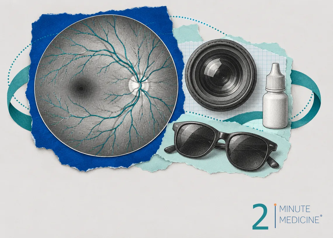
Fluorescein angiography is an eye test that uses a yellow dye and a special camera to photograph the blood vessels at the back of the eye (the retina). It shows leaking, blocked or newly grown vessels, which helps eye doctors find and treat damage from diabetes, called diabetic retinopathy. The dye goes into a vein in your arm, and the test takes about 30 minutes plus time for your pupils to widen.
Key points
- Most people with diabetes are checked with a yearly dilated eye exam and often a quick scan called OCT. Fluorescein angiography is added when the doctor needs more detail.
- It helps guide treatment, such as eye injections or laser.
- Bring sunglasses and a driver. Your vision will be blurry for several hours.
- Your skin may look yellow for a few hours, and your urine will be bright yellow or orange for a day or so.
- Brief nausea is the most common side effect. Serious allergic reactions are rare.
Diabetic retinopathy in brief
Over years, high blood sugar damages the tiny blood vessels in the retina, the layer of light-sensing tissue that lines the back of the eye. Damaged vessels can bulge, leak fluid or blood, or close off. When parts of the retina don’t get enough blood, the eye grows new vessels. These are fragile and can bleed into the eye or pull on the retina.
Leaking fluid can make the center of the retina (the macula) swell. This is called diabetic macular edema, and it’s the most common reason people with diabetes lose vision. Early retinopathy has no symptoms, so regular eye exams are the only way to catch it before sight is affected.
Why fluorescein angiography is done
Your eye doctor (an ophthalmologist or retina specialist) may order this test to:
- Find exactly where vessels are leaking before laser treatment
- Look for areas of the retina that have lost their blood supply
- Check for fragile new vessels, the sign of advanced (proliferative) retinopathy
- Explain vision loss that a regular exam doesn’t account for
- See how well treatment is working
The same test helps with other eye problems too, such as wet macular degeneration, blocked retinal veins and inflammation inside the eye (uveitis).
How fluorescein angiography works
Fluorescein is a yellow-orange dye that glows when blue light shines on it. After it’s injected into a vein in your arm, it reaches the eye within about 10 to 15 seconds. A camera with blue light and special filters takes a quick series of photos as the dye flows through the retina’s arteries and veins. Healthy vessels hold the dye inside. Damaged ones let it seep out, show up as bright spots, or stay dark where blood isn’t flowing.
Before the test
- Tell your doctor if you’ve had a reaction to fluorescein dye before, or have asthma or other allergies. A shellfish or iodine allergy isn’t a reason to avoid this dye.
- Tell them if you’re pregnant or breastfeeding. The test is usually put off during pregnancy unless it’s really needed.
- You can usually eat and take your medicines as normal, including your diabetes medicines.
- Take out contact lenses before the test.
- Bring sunglasses, and arrange for someone to drive you home.
During the test
- Drops are put in your eyes to widen (dilate) your pupils. This takes 15 to 30 minutes.
- You sit at the camera with your chin on a rest and your forehead against a bar. A few photos are taken first, without dye.
- A nurse or technician places a small needle in a vein in your arm or the back of your hand and injects the dye over a few seconds.
- The camera flashes many times over the next minute or so as the dye passes through your eye, and then takes a few more photos over the next 5 to 10 minutes.
You’ll see bright flashes and then colored afterimages for a few minutes. Some people feel warm or have a strange taste as the dye goes in. About 1 in 10 people feel sick to their stomach for a minute or two. Tell the staff if you feel itchy, faint or short of breath.
Afterward
- Vision stays blurry and bright light feels harsh for 4 to 6 hours, until the dilating drops wear off. Don’t drive until your vision is back to normal.
- Your skin may look slightly yellow for a few hours.
- Your urine will be bright yellow or orange for 24 to 36 hours as your kidneys clear the dye. Drinking water helps.
- If you’re breastfeeding, ask your doctor whether to wait before nursing, because the dye passes into breast milk.
Possible risks
Fluorescein angiography is a common, well-tested procedure. Side effects can include:
- Nausea and, less often, vomiting, which pass quickly
- A sore, yellow patch if dye leaks under the skin at the needle site. It fades over a few days. Cool compresses help.
- Itching or hives, which can be treated with an antihistamine
- A serious allergic reaction with trouble breathing or low blood pressure. This is rare, and clinics are equipped to treat it right away.
Your results
The eye doctor often reviews the photos with you the same day. In a normal test, the dye fills the vessels evenly and stays inside them. Signs of diabetic retinopathy include tiny bulges (microaneurysms), leaking dye in the macula, patches with no blood flow, and new vessels that leak heavily.
Depending on the findings, your doctor may suggest:
- Better control of blood sugar, blood pressure and cholesterol, which slows damage
- Injections into the eye of medicines that block a protein called VEGF, such as aflibercept, ranibizumab, bevacizumab or faricimab. They dry up swelling and shrink new vessels.
- Laser treatment to seal leaks or to calm the growth of new vessels
- A steroid implant placed in the eye
- Surgery (vitrectomy) to clear blood or scar tissue from inside the eye
When to call your doctor
- Pain, swelling or redness at the needle site that gets worse instead of better
- Rash or itching that starts after you get home
- New floaters, flashes of light, a dark curtain over your vision or sudden vision loss
Call 911 for trouble breathing, swelling of the face or throat, or feeling faint.
Common questions about fluorescein angiography
Is fluorescein angiography painful?
Only the needle stick, which feels like having blood drawn. The camera flashes are bright but don’t hurt, and the dilating drops may sting for a moment. Some people feel brief nausea or warmth as the dye goes in. Most people find the test easier than they expected.
What’s the difference between fluorescein angiography and OCT?
OCT (optical coherence tomography) is a quick scan with no dye or needle. It measures the thickness of the retina and shows swelling in cross-section. Fluorescein angiography shows how blood flows and where vessels leak. Doctors often use OCT routinely and add angiography when they need the flow picture.
Is there a test for diabetic retinopathy without dye?
Yes. A dilated eye exam, retinal photos and OCT can all find diabetic retinopathy without dye. Some clinics use a newer scan called OCT angiography that maps blood vessels without an injection. It shows leaks less clearly, so dye-based angiography is still used when the details matter.
Questions to ask your doctor
- Why do I need fluorescein angiography, and what are you looking for?
- What stage of diabetic retinopathy do I have?
- Do I have swelling in the macula?
- What treatment do you recommend, and how often will I need it?
- How often should my eyes be checked from now on?
Learn more
- Fluorescein angiography (MedlinePlus, U.S. National Library of Medicine)
- Diabetic retinopathy (National Eye Institute)
This guide is general information and doesn’t replace advice from your own doctor. Last updated October 2026.






 Get the app