
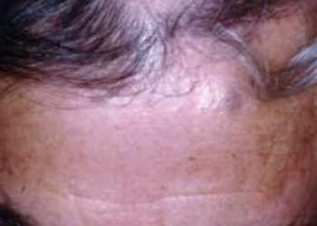
Eosinophilic infiltrates have limited diagnostic utility in alopecia areata
1. An eosinophilic infiltrate around the hair follicle was a poor diagnostic indicator of alopecia areata, especially in the chronic stage.
2. Pigmentary incontinence around the hair follicles, follicular miniaturization, and shift to the catagen or telogen phase were more useful diagnostic indicators than eosinophilic infiltrate when peribulbar lymphocytes were absent.
Evidence Rating Level: 2 (Good)
Study Rundown: Alopecia areata (AA) is a form of nonscarring hair loss believed to be caused by autoimmune destruction of the hair follicles. One of the diagnostic features of AA is the histological presence of a peribulbar lymphocytic infiltrate. This feature, however, is not always present in the chronic stage of AA. The authors of this study sought to evaluate another feature, the presence of a peribulbar eosinophilic infiltrate, for its diagnostic significance when peribulbar lymphocytes were absent. Authors found that eosinophils were a poor diagnostic feature. Among biopsies of acute stage AA, an eosinophilic infiltrate was detected in 31%, and in only 7% of those biopsies of chronic AA when a lymphocytic infiltrate was absent. The study is limited by the semi-objective nature of the histopathologic grading of the eosinophilic infiltrate.
Click to read the study in JAMA Dermatology
Relevant Reading: Histologic features of alopecia areata other than peribulbar lymphocytic infiltrates
In-Depth [prospective cohort]: This study evaluated 162 scalp biopsies taken from patients with AA for the presence of eosinophils around the hair follicles, as well as for other features such as pigmentary incontinence around the hair follicles, follicular miniaturization, and shift to the catagen or telogen phase. For control, 69 scalp specimens were obtained from patients with androgenetic alopecia, and 26 specimens from patients with trichotillomania. Lymphocytic and eosinophilic infiltrates were graded 0 (absent), 1+ (sparse to mild), 2+ (moderate), and 3+ (dense). Eosinophils were found around hair follicles in 30 of 162 specimens (18.5%) of AA in all stages. In acute stage AA, eosinophils were found in 24 of 78 specimens (30.8%) when a peribulbar lymphocytic infiltrate was present, and in chronic stage AA (where a peribulbar lymphocytic infiltrate was sparse or absent), eosinophils were found in 6 of 84 specimens (7.1%), suggesting that the presence of an eosinophilic infiltrate was of limited diagnostic usefulness. The other histologic features were found to be more useful diagnostic features in the cases of AA where a peribulbar lymphocytic infiltrate was absent: pigmentary incontinence around the hair follicles was found in 58 of 84 specimens (69.0%), follicular miniaturization in 52 (61.9%), and shift to the catagen or telogen phase in 46 (54.8%).
More from this author: Systematic reviews moderately reflect disease burden in dermatology, Bath psoralen plus ultraviolet A shows good efficacy in the treatment of mycosis fungoides, Lower rates of self skin examination among ethnic minorities
Image: PD
©2012-2014 2minutemedicine.com. All rights reserved. No works may be reproduced without expressed written consent from 2minutemedicine.com. Disclaimer: We present factual information directly from peer reviewed medical journals. No post should be construed as medical advice and is not intended as such by the authors, editors, staff or by 2minutemedicine.com. PLEASE SEE A HEALTHCARE PROVIDER IN YOUR AREA IF YOU SEEK MEDICAL ADVICE OF ANY SORT.
RelatedReports


