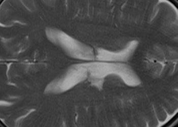
MRI may predict progression to dementia in healthy elderly adults
1. Among cognitively-intact elderly adults, magnetic resonance (MR) imaging using an arterial spin labeling (ASL) technique was shown to predict cognitive decline after 18 months of follow-up.
2. Similar ASL neuroimaging findings were identified in healthy patients that progressed to cognitive decline as compared to control subjects with known mild cognitive impairment (MCI).
Evidence Rating Level: 2 (Good)
Study Rundown: As the average life expectancy increases and the elderly population increases in size, screening for preclinical predictors of Alzheimer’s dementia has become increasingly important. Studies utilizing positron emission tomography (PET) have established that decreased metabolic activity in the posterior cingulate cortex (PCC) may be indicative of early cognitive decline, as have other biomarkers involving measurement of amyloid beta protein accumulation. Given the close relationship between perfusion and metabolism, the current study authors hypothesized that reduced perfusion, as measured by arterial spin labeling on brain MR imaging, may accurately identify patients who are at risk for impending cognitive decline. They examined a group of healthy elderly individuals, once at baseline then again after an 18-month interval, using a series of neuropsychological tests and ASL imaging. Subjects were subsequently stratified into two groups: those who displayed a decline in cognitive function and those who did not at 18 months. Results were compared to a control group with known baseline MCI. Those whose cognitive function deteriorated over the study period were found to have significantly reduced perfusion to the PCC predating their cognitive degradation as compared to those with stable cognition, a pattern similar to control subjects with known MCI. The study was designed to be highly sensitive to cognitive changes by using a low threshold for decline on neuropsychological testing, potentially limiting the specificity of the results. Additionally, ASL imaging was limited by the use of an imaging protocol that limits accuracy in patients with decreased perfusion speed. Given the complementarity of information obtained by ASL imaging and other modalities such as PET or nuclear imaging of amyloid beta protein, future studies examining the accuracy of a multimodal approach to predicting cognitive decline may yield a long-sought dementia biomarker.
Click to read the study in Radiology.
Relevant Reading: Discrimination between Alzheimer dementia and controls by automated analysis of multicenter FDG PET.
In-Depth [prospective cohort]: This study assessed baseline cognitive status by neuropsychiatric testing in a cohort of 148 healthy elderly patients initially and after an 18 month period to identify those with interval cognitive decline. All patients underwent initial brain MR imaging, including single inversion ASL imaging and calculated volumetric cerebral blood flow (CBF) mapping, normalized to gray matter volume to control for focal atrophy. Of those tested, 75 subjects (average age 73.3 years) showed no interval cognitive decline while 73 subjects (average age 73.7 years) displayed cognitive deterioration, as assessed by neuropsychiatric test performance of at least 0.5 standard deviations below baseline. These subgroups were subsequently compared to a control group of 63 patients with known MCI who underwent similar imaging and neuropsychiatric testing. In the control group, reduced CBF was observed primarily in the PCC, a region associated with default mode network activity, and was identified by unbiased whole-brain perfusion analysis. The same hypoperfusion pattern was observed at baseline in those subjects who developed subsequent cognitive impairment, but was not seen in those who remained cognitively stable (P < 0.001). A perfusion threshold of 58.5 mL/100 g/min in the PCC was used to discriminate subjects who were or were not at risk for developing subsequent cognitive decline with a sensitivity and specificity of 58.9% and 65.3%, respectively.
More from this author: Preoperative MRI improves surgical outcomes in robotic prostatectomy, Many lung cancers visible on prior imaging studies, Perfusion imaging may be useful for selecting stroke patients for thrombolysis.
Image: PD
©2012-2014 2minutemedicine.com. All rights reserved. No works may be reproduced without expressed written consent from 2minutemedicine.com. Disclaimer: We present factual information directly from peer reviewed medical journals. No post should be construed as medical advice and is not intended as such by the authors, editors, staff or by 2minutemedicine.com. PLEASE SEE A HEALTHCARE PROVIDER IN YOUR AREA IF YOU SEEK MEDICAL ADVICE OF ANY SORT.


