

Patient Basics: Dislocated Lens
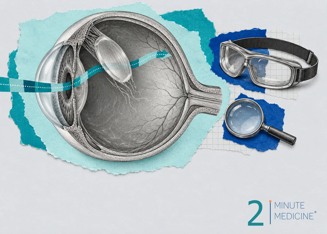
A dislocated lens (lens dislocation, or ectopia lentis) happens when the clear lens inside the eye shifts out of its normal place behind the pupil. It can slip partway or come loose completely. The most common signs are blurry vision and double vision in one eye. An eye injury is the usual cause, but a lens that moves without an injury can point to an inherited condition such as Marfan syndrome, so it is worth a full checkup.
Key points
- The lens is held in place by hundreds of tiny fibers (zonules). When they stretch or break, the lens moves.
- Causes include a blow to the eye, aging changes, past eye surgery and inherited conditions.
- An artificial lens put in during cataract surgery can also shift, sometimes years later.
- Glasses or contact lenses are enough for some people. Others need surgery to remove or reposition the lens.
- Sudden eye pain, redness and blurred vision need emergency care. A loose lens can block fluid and raise eye pressure.
How the lens normally stays in place
The lens sits just behind the colored part of the eye (the iris). It focuses light onto the retina at the back of the eye. A ring of thin fibers holds it in the middle, like the springs around a trampoline. If some fibers fail, the lens tilts or slides to one side. Doctors call this a partial dislocation (subluxation). If all the fibers fail, the lens can fall back into the gel that fills the eye or move forward toward the cornea.
What causes a dislocated lens
- Eye injury. A hard blow from a ball, fist, fall or car crash is the most common cause. Damage may show up right away or months later.
- Marfan syndrome. This inherited connective tissue disorder is the most common genetic cause. The lens usually shifts upward and outward, and it often happens in both eyes.
- Homocystinuria. In this rare inherited problem with processing protein, the lens tends to drop downward. It also raises the risk of blood clots.
- Other inherited conditions, such as Weill-Marchesani syndrome and some types of Ehlers-Danlos syndrome, or a lens shift that runs in a family on its own
- Pseudoexfoliation syndrome. In this age-related condition, flaky material builds up inside the eye and weakens the fibers.
- Other eye problems, including very high nearsightedness, a very mature cataract, long-term eye inflammation, past eye surgery, and some cases of glaucoma or retinitis pigmentosa
Dislocated intraocular lens
During cataract surgery, the cloudy natural lens is replaced with a clear plastic one (an intraocular lens, or IOL). It sits in the thin pouch that held the old lens. Years later, the pouch’s support fibers can weaken, and the whole lens and pouch can slip down. This is more likely with pseudoexfoliation, a past eye injury or earlier retina surgery.
Dislocated lens symptoms
- Blurry vision that glasses no longer fix
- Double vision in one eye. You still see two images when you cover the other eye.
- Sudden changes in your glasses prescription, often more nearsighted
- Glare, or seeing the curved edge of the lens
- Vision that seems to shake or shift when you move your eye
- Sudden pain, redness, headache, halos around lights and nausea, if the lens blocks fluid drainage and pressure rises
A slight shift may cause no symptoms and be found at a routine eye exam. Children may not complain at all, so a failed school vision screening or squinting can be the first clue.
How it’s diagnosed
An eye doctor (ophthalmologist) examines the eye with a slit lamp, a microscope with a bright light, after drops widen the pupil. They can often see the lens edge or watch the lens and iris wobble when the eye moves. They also check your eye pressure and look at the retina. If bleeding or a cloudy lens blocks the view, an ultrasound of the eye helps.
When a lens moves without a clear injury, especially in a child or young adult, the doctor looks for a cause. This may mean a heart ultrasound and a genetic test for Marfan syndrome, or a blood or urine test for homocystinuria. Finding these conditions early protects the heart and blood vessels, and it matters before any surgery.
How a dislocated lens is treated
Without surgery
If the lens has shifted only a little and vision is still good, glasses or contact lenses may correct it. Some people see better through the part of the pupil that no longer has a lens in front of it, and special glasses can help with that. Children need close follow-up and sometimes patching of the stronger eye to prevent a lazy eye (amblyopia).
Surgery
Surgery is usually advised when:
- Glasses can’t give useful vision
- The lens has moved forward or is pressing on the iris and raising eye pressure
- The lens is causing inflammation or has a cataract
- The lens is fully loose inside the eye
The surgeon removes the loose lens, often along with some of the gel inside the eye (vitrectomy). A new artificial lens is then placed in front of the iris, clipped to the iris, or held by stitches or tiny anchors in the white wall of the eye. For a dislocated intraocular lens, the surgeon may reposition and secure the same lens or swap it for a new one. Retina specialists and surgeons who focus on the front of the eye usually do these repairs.
Recovery
Most people go home the same day. You will use eye drops for several weeks, may wear a shield at night, and should avoid heavy lifting and rubbing the eye. Vision often improves over a few weeks. Possible problems include higher eye pressure, swelling in the retina, infection and retinal detachment.
Lowering your risk
- Wear protective eyewear for sports such as racquetball and basketball, and for yard work and power tools.
- If you have Marfan syndrome or another condition linked to lens problems, see an eye doctor every year.
- Keep regular eye exams after cataract surgery, especially if you have pseudoexfoliation.
When to see a doctor
Make an eye appointment soon for new blurry vision, double vision in one eye or a sudden change in how your glasses work.
Get emergency care for:
- Sudden eye pain with redness, halos, headache or vomiting
- A hard hit to the eye followed by any vision change
- New flashes of light, a shower of floaters, or a shadow or curtain over part of your vision
Common questions about lens dislocation
Can a dislocated lens be fixed?
Yes. Many people regain good vision. A slightly shifted lens can often be managed with glasses or contacts. A badly shifted lens is removed and replaced with an artificial lens secured in a new position. A dislocated implant from cataract surgery can usually be repositioned or exchanged.
Is a dislocated lens the same as a dislocated eyeball?
No. A dislocated lens happens inside the eye, and the eye looks normal from the outside. In a dislocated eyeball (globe luxation), the whole eye bulges forward past the eyelids. That is rare and needs emergency care. A broken eye socket is a different injury too.
Why do I have double vision in one eye?
Double vision that stays when you cover the other eye comes from a problem inside that eye. A shifted lens is one cause. Cataracts, dry eye, astigmatism and corneal problems are more common causes. An eye doctor can usually find the reason with a standard exam.
Questions to ask your doctor
- How far has my lens moved, and is it still moving?
- Can glasses or contacts work for me, or do I need surgery?
- Should I be tested for Marfan syndrome or another condition?
- What kind of replacement lens would you use, and how is it held in place?
- What signs after surgery mean I should call right away?
Learn more
- Isolated ectopia lentis (MedlinePlus Genetics, U.S. National Library of Medicine)
- Eye diseases (MedlinePlus, U.S. National Library of Medicine)
This guide is general information and doesn’t replace advice from your own doctor. Last updated October 2026.






 Get the app