No products in the cart.

Patient Basics: X-Rays
Originally published by Harvard Health.
What Is It?
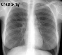
X-rays are waves of electromagnetic radiation that are used to create images of organs and other structures inside the body. X-rays have a very short wavelength. As they penetrate the body, they are absorbed in different amounts by different body tissues. For example, bones are dense and absorb X-rays very well, but soft tissues (skin, fat, muscle) allow more X-rays to pass through. The result is an X-ray shadow on a film or fluorescent screen, where images of bones appear white, while shadows of soft tissues appear as various shades of gray.
In some forms of X-rays, a chemical called contrast medium is given to the patient to help outline a specific body area on X-ray film. This chemical can be swallowed, given as an enema or injected into a vein. The contrast medium appears white on the X-ray film, and can produce a sharp outline of structures such as the digestive tract and the paths of blood vessels.
While X-rays themselves are painless, there may be some mild discomfort from a pin prick or from an enema if contrast medium is used. Some X-rays take less than a minute, while longer X-ray procedures may take an hour or more.
What It’s Used For
X-rays are used for many purposes, including determining if a bone is broken, seeing whether an internal organ is infected, and looking for cancer. There are many different types of X-rays currently used to detect cancer. For example, both mammography (a series of breast X-rays) and the barium enema (a series of bowel X-rays with contrast medium), are routine procedures sometimes used for cancer screening in adults of certain age groups. To check for tumors in precise cross-sections of the body, a computed tomography (CT) scan also can be used. A CT scan is a series of X-rays linked to computer technology. Even without using specialized techniques, uncomplicated, routine X-rays often can show abnormal shadows or silhouettes that might be cancerous tumors.
Preparation
There are many different types of X-ray procedures, and some require special preparation. For example, before having X-rays of your digestive tract, you may need to change your diet, fast entirely or use laxatives or enemas. Before having mammography, you must avoid using deodorants, body powders, perfumes and body creams, which can produce abnormal shadows in the mammogram image. You should remove all jewelry from the part of your body to be X-rayed.
X-rays can affect a developing fetus. If you are a woman and there is a chance that you might be pregnant, tell your doctor before you have an X-ray.
How It’s Done
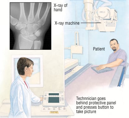
You likely will be asked to remove your clothing over the part of your body to be X-rayed. You will be given a hospital gown. For certain X-ray procedures, you also will be given a flexible lead apron or other type of protective drape to shield portions of your body from unnecessary X-ray exposure. You will be asked to either stand on the floor or lie or sit on a table in an X-ray room, and a technician will position your body in a way that gives the best X-ray view.
The technician will position the X-ray machine near your body, so that the machine’s X-ray tube (where the X-rays come out) is pointing at the correct body area. After going behind a protective panel, the technician will press a button to take the X-ray picture.
For more specialized series of X-rays, such as mammography or a CT scan, the procedure is slightly more complicated.
Follow-Up
Your X-rays will be read by a radiologist who will report the result to your doctor. Call your doctor’s office for the official X-ray report.
Risks
Although large doses of X-ray radiation are harmful, modern X-ray facilities use techniques and equipment that keep your X-ray exposure to a minimum. Lead aprons and other types of lead shields also can be used to protect your reproductive organs and other parts of your body during the X-ray procedure.
In general, a pregnant woman should avoid X-rays because of risks to her unborn child. Also, when growing children are scheduled for X-rays, parents should make sure that these tests are really necessary, that there are no alternative procedures that don’t use radiation (such as magnetic resonance imaging (MRI) or ultrasound), and that as little of their child’s body as possible is exposed during the X-ray session.
Over a lifetime, you can help minimize your X-ray exposure by keeping track of where and when you had X-rays in the past, and by telling your doctor about your previous films when appropriate. In some cases, this will help you to avoid having duplicate X-rays. Since many X-ray facilities destroy their films after 7 to 10 years, you may want to ask for your old X-rays to keep for your records.
When To Call a Professional
Routine diagnostic X-rays usually do not cause any side effects. However, if you received an injection of contrast medium before your X-rays, call your doctor if you have bleeding, pain, swelling or redness at the injection site. Ask your doctor about other signs or symptoms to watch for after your specific X-ray procedure.
Additional Info
American College of Radiology
1891 Preston White Drive
Reston, VA 20191
Toll-Free: 1-800-227-5463
http://www.radiologyinfo.org/
Radiological Society of North America
820 Jorie Blvd.
Oak Brook, IL 60523-2251
Phone: 630-571-2670
Toll-Free: 1-800-381-6660
http://www.radiologyinfo.org/
National Library of Medicine (NLM)
8600 Rockville Pike
Bethesda, MD 20894
Phone: 301-594-5983
Toll-Free: 1-888-346-3656
http://www.nlm.nih.gov/
U.S. Food and Drug Administration (FDA)
5600 Fishers Lane
Rockville, MD 20857
Toll-Free: 1-888-463-6332
http://www.fda.gov/

