
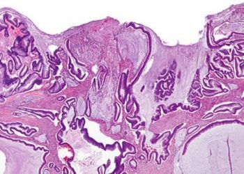
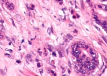
CT colonography may help distinguish chronic diverticular disease from cancer
1. In the evaluation of focal mass-like findings in the sigmoid colon on computed tomographic (CT) colonography, segmental bulging and the absence of diverticula strongly predicted carcinoma over chronic diverticular disease.
Evidence Rating Level: 3 (Average)
Study Rundown: Computed tomographic (CT) colonography is a non-invasive method of evaluating for polyps and cancers within the colon, and the results of prior studies support its utility as an adjunct to optical colonoscopy in colorectal cancer screening. However, carcinoma of the sigmoid colon has been previously shown to share similar CT imaging features with chronic diverticular disease. Given the significant difference in management of these two conditions, accurate differentiation of carcinoma from diverticular disease may help reduce under-diagnosis and over-treatment. The purpose of this trial was to identify predictive morphologic features of carcinoma in the evaluation of sigmoid focal mass-like findings using CT colonography. Images from patients with focal abnormalities in the sigmoid colon on CT colonography were retrospectively evaluated for the presence of several previously-described imaging features. The authors demonstrated that the absence of diverticula within the affected segment and bulging of the proximal or distal ends of the segment—the “shoulder phenomenon”—were most predictive of carcinoma as compared to chronic diverticular disease. The presence of colonic wall thickening or pericolonic fat infiltration were not demonstrated to be predictive for sigmoid carcinoma. None of the evaluated features were shown to be predictive of carcinoma stage or the presence of mass infiltration beyond the colonic wall. The main limitations of this study were the retrospective methodology and the fact that the evaluators were unblinded to the final diagnoses. Further prospective trials are needed to validate these results in a larger patient cohort and to determine the utility of using specific morphologic features in improving patient outcomes.
Click to read the study in Radiology
Relevant Reading: Current status on performance of CT colonography and clinical indications
In-Depth [retrospective cohort]: This study retrospectively analyzed consecutive patients who underwent CT colonography at two non-academic centers in the Netherlands from 2007 to 2012. Focal sigmoid lesions were defined as the presence of abnormal sigmoid segment length of >20mm, bowel wall thickening >5mm, and distorted mucosal fold patterns on CT colonography. Final diagnoses were confirmed by surgery, endoscopy, or negative clinical follow-up two years after CT colonography. Overall, 212 patients (chronic diverticular disease=97, sigmoid carcinoma=115) with focal findings in the sigmoid colon were included. The presence or absence of specific imaging features was determined via consensus by four independent, unblinded readers. The absence of diverticula on imaging demonstrate a positive predictive value (PPV) of 93% and a negative predictive value (NPV) of 95% for sigmoid carcinoma. The presence of shouldering phenomenon demonstrated a PPV of 75% and a NPV of 92%. The combination of both features yielded a diagnostic accuracy of 93% (95% CI: 82-95%) for sigmoid carcinoma.
More from this author: No association found between time to appendectomy and appendix perforation, Obesity linked with prostate cancer progression, Use of 5α-reductase inhibitors may not increase risk of high-grade prostate cancer
Image: PD
©2014 2 Minute Medicine, Inc. All rights reserved. No works may be reproduced without expressed written consent from 2 Minute Medicine, Inc. No article should be construed as medical advice and is not intended as such by the authors, editors, staff or by 2 Minute Medicine, Inc.


