
Lumicell: Injectable imaging agent illuminates residual breast cancer tissue during surgery to ensure tumor removal.
- In the intervention group of 357 patients, 27 had additional removal of residual tumor based on imaging agent-guided technology.
- Six of 357 patients had adverse events and no deaths were reported in this trial.
The Latest
A recent multi-center phase 3 clinical trial funded by Lumicell investigated the safety and efficacy of LUM Imaging System in identifying residual cancer in lumpectomy beds of female breast cancer patients during surgery. Four hundred patients were injected with the LUM015 imaging agent which emits a fluorescent signal upon interacting with cancerous cells that can be visualized using the LUM Imaging system. Patients underwent lumpectomy and were then randomized in a 1:10 ratio of control vs. intervention. Patients in the intervention group underwent additional LUM015-guided evaluation for residual tumor. Of 357 patients in the intervention group, 27 underwent removal of additional tumor left behind after standard lumpectomy. Second surgeries were avoided by LUM015 in 9 of 62 patients with positive margins. On per-margin analysis, LUM015 had 85.2% specificity and 49.3% sensitivity. Six patients had adverse events and no deaths were reported in this trial.
Physicians Perspective
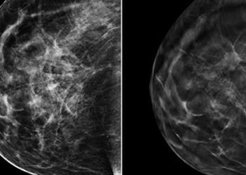
Breast cancer is one of the most common malignancies in women worldwide. Although breast-conserving surgery (lumpectomy) provides equivalent survival for patients compared to mastectomy, local recurrence after lumpectomy increases cancer mortality. One of the strongest predictors of local recurrence is positive lumpectomy margins, which suggest incomplete tumor removal. Currently, examination of lumpectomy margin usually occurs days after surgery and are often positive in 20-40% patients, with some needing re-excision surgery. This imaging technique provides a novel method to survey the entire lumpectomy cavity intraoperatively and allow immediate identification and removal of any residual tumor.
Molecular Target of Therapy
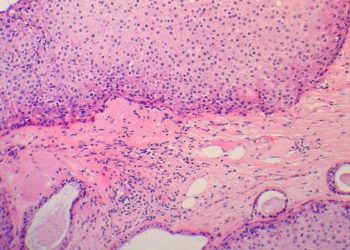
As mentioned, Lumicell, in collaboration with Mass General Hospital developed a novel injectable PEGylated protease-activated far-red fluorescent imaging agent. This agent becomes optically active when cleaved by proteases that are overexpressed by tumors. The probe in the LUM optical head can excite LUM015 and collect real-time fluorescent recordings of the lumpectomy cavity.
Company History
Lumicell’s fluorescent visualization system was initially developed in the lab of co-founder Moungi Bawendi at Massachusetts Institute of Technology. The current injectable therapy was developed by Lumicell in collaboration with Duke University and Massachusetts General hospital. Lumicell received FDA approval for both the imaging agent and the direct visualization system in 2024 and has ongoing studies for applying their technology in gastrointestinal tumors and sarcomas.
Further reading: https://evidence.nejm.org/doi/10.1056/EVIDoa2200333
©2024 2 Minute Medicine, Inc. All rights reserved. No works may be reproduced without expressed written consent from 2 Minute Medicine, Inc. Inquire about licensing here. No article should be construed as medical advice and is not intended as such by the authors or by 2 Minute Medicine, Inc.
RelatedReports


