Patient Basics: Detached Retina
Originally published by Harvard Health.
What Is It?
The retina is the light-sensitive layer at the back of the eye that converts light images into nerve impulses that are relayed to the brain to produce sight. When the retina separates from the deeper layers of the eyeball that normally support and nourish it, the retina is said to be detached. Without this nourishment and support, the retina does not function properly, and this can cause a variety of visual symptoms. For example, if the retina detaches near the macula, the part of the eye that is responsible for the center of the visual field (reading for instance), then there may be a sudden, significant blurring or loss of vision. However, if the area of detachment is closer to the outer edges of the retina, then the visual loss may be more like a curtain being drawn over one side of the visual field (the “curtain effect”). Other symptoms of retinal detachment may include floating shapes in the field of vision or brief flashes of light.
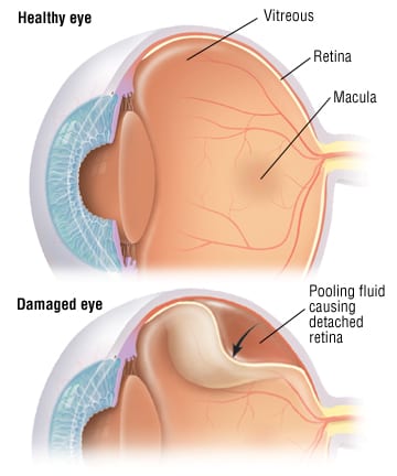
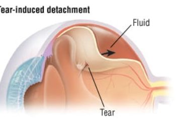
Although there are several types of retinal detachment, the most common one starts when a tear or hole develops in the retina, and some of the gel-like substance that fills the inside of the eye (vitreous fluid) leaks through the opening. Eventually, the leaking vitreous fluid gets behind the retina, separating the retina from other layers of the eye.
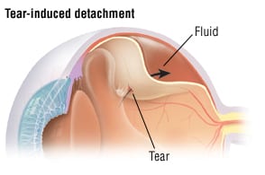
The retinal tear that triggers a retinal detachment sometimes is caused by trauma. More often, it is caused by a change in the gel-like consistency of the vitreous fluid that can occur as a part of aging. This age-related change can occur unpredictably in older people, and there is no way to prevent it. Once this type of retinal detachment develops and causes symptoms, it can progress to total blindness if it is not repaired promptly.
In the United States, retinal detachment is a relatively uncommon condition, affecting only one of every 10,000 Americans each year. However, there are certain groups who have an unusually high risk of developing this problem, including:
- People who have had cataract surgery — Up to 3% of people who have had cataract surgery eventually develop a detached retina, making retinal detachment the most serious post-surgical complication of cataract treatments. The high rate of retinal detachments after cataract surgery may be related to the vitreous fluid inside the eye becoming watery instead of gel-like after surgery. If there has been a complication at the time of cataract surgery especially if the posterior capsule of the human lens has torn and there has been loss of the vitreous, jelly retinal complications especially detachment is even more common.
- Severely nearsighted people — These people often are nearsighted because they have an eyeball that is unusually long. The elongated shape creates more stress between the vitreous fluid and the surface of the retina.
- People who have had blunt trauma to the eye or penetrating eye injuries
In many cases, the more risk factors a person has, the higher the risk of retinal detachment. For example, a very nearsighted person who also undergoes cataract surgery probably will have a higher risk of retinal detachment than someone who has cataract surgery and is not nearsighted. Overall, the risk of retinal detachment increases the older you get, and men are about 50% more likely to develop the problem than women.
People with diabetes also have a higher risk of a different type of retinal detachment as a complication of diabetic retinopathy, a family of retinal disorders thought to be related to abnormal or erratic blood sugar levels.
Symptoms
Symptoms of a detached retina may include:
- The sudden appearance of “floaters” (dark, semi-transparent, floating shapes) in the field of vision. Most worrisome is a shower of black dots. These are actually red blood cells because all retinal tears bleed a little when they occur.
- Brief, bright flashes of light — These flashes may be most noticeable when you move your eyes in the dark.
- Loss of central vision
- Loss of peripheral vision (the curtain effect)
Diagnosis
After reviewing your symptoms, your doctor will ask about your medical history, especially any history of cataract surgery, nearsightedness, and diabetes or eye trauma. Next, your doctor will examine your eye, and will test how well you can see and your peripheral vision (side vision). Finally, the doctor will use special eye drops to dilate (open) your pupils so he or she can examine the inside of your eye, including the retina. During this retinal examination, the doctor will check for retinal tears and areas of detachment by using a special hand-held ophthalmoscope (a lighted instrument for looking inside the eye) or a slit lamp. If necessary, your doctor will do more tests to determine the extent of your visual field loss.
In cases in which the doctor cannot see a retinal detachment while examining your eye, an ultrasound of the eye may be necessary. This can occur when the pupil cannot be fully dilated or there is some cloudiness inside the eye that prevents the doctor from seeing the retina.
Expected Duration
Once a retinal detachment develops and causes symptoms, it should be treated quickly to preserve as much vision as possible. Ideally, the repair should be done less than one week after the detachment began. For detachments threatening the macula (central vision), it is best to treat the problem within the first 24 hours.
Prevention
Most retinal detachments are age related, and there is no way to prevent them. If you are middle-aged or older, you may be able to identify eye problems in their early stages by scheduling an eye examination with an ophthalmologist every one to two years.
To prevent retinal detachments that are caused by diabetic retinopathy, people with diabetes should try to maintain near-normal blood sugar levels and to avoid wide swings between high and low blood sugar levels. Also, all diabetics should have a thorough eye examination through dilated pupils at least once a year. If any retinopathy is detected the examinations should be done every six months.
Treatment
If you have a detached retina, you should be treated by an ophthalmologist, a physician who specializes in eye problems. Several techniques are available to repair retinal tears and to eliminate the area of separation behind the detached retina. Some options include (in some cases more than one of the options listed below may be combined):
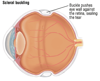
- Scleral buckling — First, a tiny hole is made in the sclera (the tough layer beneath the retina, also known as the white of the eye). Through this tiny hole, any vitreous fluid that has leaked behind the retina is drained, allowing the detached retina to fall back into its normal position. Next, a small tuck or indentation is made in the sclera and secured with a silicone buckle.
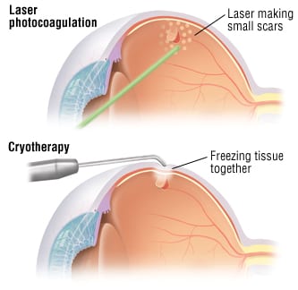
- Cryotherapy — The retinal tear is sealed with a freezing probe.
- Laser photocoagulation — A laser beam is focused on the retinal tear to seal it.
- Pneumopexy — A bubble of special gas is injected near the area of retinal detachment to press the retina back into place.
- Vitrectomy — Part of the vitreous fluid is removed near the detachment and then replaced with sterile saline (a salt solution) or some other fluid.
Once treatment is complete, you will need to return to your ophthalmologist for regular follow-up visits. These visits are necessary to check for signs that the retina has detached again in your treated eye or if the problem is happening in your untreated eye. People who already have had a retinal detachment in one eye have an increased risk of detachment in the other eye.
When To Call a Professional
Call your doctor immediately if you have symptoms of a detached retina, especially if you have a history of cataract surgery, severe nearsightedness, eye trauma, diabetes, or previous treatment for a detached retina.
Prognosis
With proper treatment, the prognosis is excellent. More than 90% of detached retinas can be reattached successfully. In some cases, more than one treatment is necessary.
Vision is most likely to return to near normal if the problem is treated less than seven days after the detachment begins. Some blurring of vision may remain in people who have detachments that involve the macula (central vision). This is why treatment is an emergency if the macula is still attached.
Additional Info
National Eye Institute
2020 Vision Place
Bethesda, MD 20892-3655
Phone: 301-496-5248
http://www.nei.nih.gov/
National Institute on Aging
Building 31, Room 5C27
31 Center Drive, MSC 2292
Bethesda, MD 20892
Toll-Free: 800-222-2225
http://www.nih.gov/nia/
American Academy of Ophthalmology
P.O. Box 7424
San Francisco, CA 94120-7424
Phone: 415-561-8500
Fax: 415-561-8533
http://www.aao.org/