
Thrombolysis harmful in acute ischemic strokes with large area of parenchymal hypoattenuation [Classics Series]
This study summary is an excerpt from the book 2 Minute Medicine’s The Classics in Medicine: Summaries of the Landmark Trials
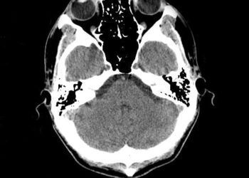
1. Parenchymal hypoattenuation of greater than 33% of the middle cerebral artery (MCA) territory on computed tomographic (CT) imaging within 6 hours of onset of an acute stroke was predictive of poor neurologic outcomes with intravenous thrombolytic therapy (tPA) and an increased risk of hemorrhagic conversion.
2. Like those with a large area of focal hypoattenuation, patients without parenchymal abnormalities on CTshowed a significantly increased risk of cerebral hemorrhage and no significant change in neurologic outcomes at 90-days post-tPA; however, those with small (< 33% of MCAterritory) regions of hypoattenuation showed a significant improvement in long-term function.
Original Date of Publication: November 1997
Study Rundown: Acute ischemic stroke occurs when blood supply to a region of the brain is disrupted, leading to characteristic symptomatology related to the location of the lesion. After clinical examination, the mainstay of stroke diagnostics depends upon neuroimaging to aid in lesion localization and characterization of its type, size, and the extent of irreversible ischemia to guide therapeutic intervention. While recent trials have established mechanical thrombectomy as the optimal therapeutic strategy in large-vessel occlusions, the initial management of ischemic strokes presenting within 3-6 hours of onset was previously established by the ECASS and NINDS trials, which revealed that early tPA administration may achieve chemical recanalization and improve neurologic recovery. Both modern directed intra-arterial therapies and standard intravenous tPA administration are predicated on the concept that the affected tissue has not progressed to complete infarction, or that there is a region of hypoperfused or “penumbral” tissue within the affected vascular territory that may be rescued by the restoration of blood flow. The referenced study sought to determine if a large area of hypoattenuation on initial CT, indicative of terminal ischemic edema, may portend poorer outcomes with standard tPA treatment. Patients presenting with acute ischemic stroke within 6 hours of symptom onset underwent baseline head CT and were randomized to receive either tPA or placebo. Patients were then prospectively subcategorized into one of three groups according to the extent of visible MCA territorial hypoattenuation: no visible defects, 33% or less, or more than 33% of the region affected, and neurologic outcomes were recorded at 90 days post-treatment. The authors found that those with greater than 33% of the MCA territory visibly hypoattenuated and those with no visible hypoattenuation on initial CT imaging did not have a statistically significant improvement in long-term neurologic recovery, but did suffer a significantly increased risk of fatal cerebral hemorrhage secondary to tPA therapy. Those with hypoattenuation visible in less than 33% of the MCA territory were not at significantly increased risk of hemorrhagic conversion and demonstrated an improvement in long-term neurologic recovery, as observed in the previously referenced trials. This analysis was instrumental in demonstrating that the extent of completed ischemia visible on CT could predict outcomes after stroke therapy.
Click to read the study in Radiology
In-Depth [randomized controlled trial]: A total of 620 patients presenting with acute, ischemic hemispheric stroke within 6 hours of symptom onset were randomized to either receive intravenous tPA or placebo in a double-blinded fashion across multiple European medical centers. All patients underwent baseline head CT, and were prospectively categorized into one of three groups according to the extent of hypoattenuation visible within the affected MCA territory: no hypoattenuation visible, less than 33% (small territory), or greater than 33% (large territory.) Of those enrolled, 336 patients had no visible parenchymal hypoattenuation, while 215 displayed a small region, and only 52 displayed a large region of hypoattenuation, and no significant differences in baseline characteristics were noted between the subcategories except for their presenting clinical stroke scores (progressively worsened for those with larger areas of infarction; p < 0.0001.) The degree of presenting hypoattenuation was significantly associated with an increased baseline risk of poor neurologic outcomes as assessed within the placebo-treated groups (p < 0.0001). Among patients treated with intravenous tPA, no statistically significant effect on long-term neurologic recovery was noted for patients with either no initial parenchymal hypoattenuation or those with a large area of parenchymal hypoattenuation. However, 14 of 198 patients (7%) in these two groups died secondary to cerebral hemorrhage, while only 2 of 190 patients (1%) in the corresponding placebo-treated groups suffered the same complication. Patients who presented with a small area of hypoattenuation displayed an OR for good neurologic recovery of 3.43 following tPA treatment, while those with a large or no area of hypoattenuation did not show a significant difference in their odds of recovery (OR 0.41, 95%CI 0.06-2.70; OR 1.27, 95%CI 0.82-1.95, respectively.) This study established the lack of benefit and subsequent risks of utilizing intravenous thrombolytic therapy in patients with greater than one-third of the MCA territory visibly infarcted.
Von Kummer R, Allen KL, Holle R, Bozzao L, Bastianello S, Manelfe C, et al. Acute stroke: Usefulness of early CT findings before thrombolytic therapy. Radiology. 1997 Nov 1;205(2):327–33.
Hacke W, Kaste M, Fieschi C, Toni D, Lesaffre E, Kummer R von, et al. Intravenous Thrombolysis With Recombinant Tissue Plasminogen Activator for Acute Hemispheric Stroke: The European Cooperative Acute Stroke Study (ECASS). JAMA. 1995 Oct 4;274(13):1017–25.
The National Institute of Neurological Disorders and Stroke rt-PA Stroke Study Group. Tissue plasminogen activator for acute ischemic stroke. The New England Journal of Medicine. 1995 Dec 14;333(24):1581–87.
©2022 2 Minute Medicine, Inc. All rights reserved. No works may be reproduced without expressed written consent from 2 Minute Medicine, Inc. Inquire about licensing here. No article should be construed as medical advice and is not intended as such by the authors or by 2 Minute Medicine, Inc
RelatedReports

![siRNA against antithrombin alleviates symptoms of hemophilia [PreClinical]](https://www.2minutemedicine.com/wp-content/uploads/2015/04/clot-CCWiki-350x250.jpg)


