


Frequency of pulmonary embolism amongst patients admitted for first-time syncope
1. Pulmonary embolism was found in 17.3% of patients admitted for first episodes of syncope.
2. For admitted patients with no alternative explanation for syncope, 25.4% (52/205) were found to have a pulmonary embolus.
Evidence Rating: 2 (Good)
Study Rundown: Syncope is a short term loss of consciousness due to lack of cerebral perfusion often caused by neurologic or cardiovascular mechanisms. Pulmonary embolism is considered to be one of the potential causes for syncope, though a diagnostic workup is rarely performed for patients hospitalized for syncope. Because of this, little is known about the how common pulmonary embolism is for these patients. This study looked to assess all patients for pulmonary embolism who were admitted for a first episode of syncope in order to gauge prevalence.
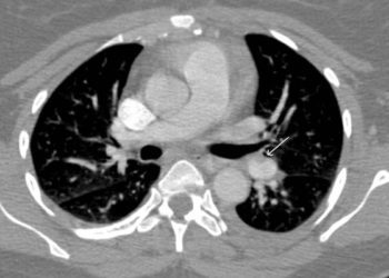
All patients admitted to an inpatient service for first-time syncope from the emergency departments of 11 Italian hospitals from 2012-2014 were evaluated for pulmonary embolism. For the 560 patients who met study inclusion criteria, pulmonary embolism related histories and D-dimer assays were obtained. Based on these factors, patients were categorized as “likely” or “unlikely” to have a pulmonary embolism. Those in the “likely” group were imaged with computed tomographic pulmonary angiography (CT scan) or ventilation-perfusion lung scanning (VQ scan) to determine if a pulmonary embolism was present. Of the 560 study patients, 230 (41%) were in the “likely” group and evaluated for pulmonary embolism. Pulmonary embolisms were found in 40% (72/180) of patients getting a CT scan, 49 % (24/49) of patients getting VQ scans, and in the autopsy of a patient who passed away (1/1). Patients with no explanation for syncope were found to have a pulmonary embolus 25.4% (52/205) of the time.
Click to read the study, published today in NEJM
Relevant Reading: Acute pulmonary embolism
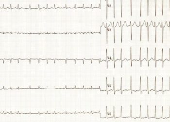
In-Depth [cross-sectional study]: This study was conducted from March 2012 to October 2014 at 11 Italian hospitals. Patients seen in the emergency department (n = 2584) and subsequently admitted to the hospital (n = 717) were considered for the study. Reasons for admission were often failure to identify cause of syncope, fall related trauma, or high suspicion of cardiac related cause of syncope. Patient workup included Wells criteria questions for determining pretest probability of pulmonary embolism, D-dimer assay, chest X-ray, electrocardiogram, and routine blood testing. If indicated, patients received prophylactic anticoagulation. Based on patient history and D-dimer results, patients included in the study (n = 560) were classified as “likely” of “unlikely” to have a pulmonary embolism. No further pulmonary embolism workup was done for the “unlikely” group (n = 330, 59%), while those in the “likely” group (n = 230, 41%) were scanned with computed tomographic pulmonary angiography or VQ regimen as clinically appropriate. Pulmonary embolism was determined if CT scan showed an intraluminal filling defect or a VQ scan showed a perfusion defect of >75% of a normally ventilated segment. Pulmonary embolism was detected on 40% of CT scans (72/180), 49% of VQ scans (24/49), and in one autopsy performed (1/1). Overall, pulmonary embolism was detected in 17.3% (97/560) of patients in the study. On CT scans, emboli were located in the main pulmonary artery (30/72), lobar arteries (17/72), segmental arteries (19/72), and subsegmental arteries (5/72). Of patients presenting with syncope of undetermined origin (n = 205), pulmonary embolism was detected for 52 (25.4%; 95%CI 19.4 to 31.3). For 355 patients whose syncope was thought to be from another cause, pulmonary embolism was detected in 45 (12.7%; 95%CI 9.2 to 16.1).
Image: CC/Wiki
©2016 2 Minute Medicine, Inc. All rights reserved. No works may be reproduced without expressed written consent from 2 Minute Medicine, Inc. Inquire about licensing here. No article should be construed as medical advice and is not intended as such by the authors or by 2 Minute Medicine, Inc.
RelatedReports