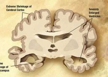
Amyloid may cause distant cerebral hypometabolism in Alzheimer’s
1. In a longitudinal study of 15 patients with Alzheimer’s disease (AD), regions of isolated cerebral hypometabolism detected by FDG-PET correlated with increase in amyloid deposition in anatomically separate but functionally connected brain regions.
Evidence Rating Level: 3 (Average)
Study Rundown: The current understanding of Alzheimer’s disease (AD) pathophysiology involves deposition of amyloid plaques resulting in localized cerebral inflammation and toxicity. Previous work using position emission tomography (PET) of the brain in patients with AD using radioligands for metabolism (fludeoxyglucose, or 18F-FDG) and amyloid plaques (Pittsburg Compound B, or 11C-PiB) have shown strong correlation between the area of hypometabolism and high amyloid deposition. However, FDG-PET has also demonstrated that AD brains contain areas in which brain hypometabolism occur without high-amyloid burden. The purpose of this trial was to explore the hypothesis that these hypometabolism-only (HO) areas are related to amyloid toxicity of anatomically remote, but functionally related areas of the brain. The authors prospectively evaluated 15 patients with AD with baseline and follow-up PET examinations using 18F-FDG and 11C-PiB over a span of two years and correlated their findings with internal functional connectivity maps as determined by resting-state functional MRI (rs-fMRI). At the conclusion of the trial, the authors found a significant positive correlation between the longitudinal changes in metabolism of the HO-areas with remote functionally interconnected brain regions. Furthermore, the authors reported a significant correlation between the loss of metabolism in the HO-regions and clinical cognitive decline. The results of this study support the hypothesis that a decrease in brain metabolism can decrease as a result of amyloid toxicity of remote, but functionally connected regions. However, the study is limited by the small sample size; larger studies are required to accurately describe this relationship.
Click to read the study in Journal of Nuclear Medicine
Relevant Reading: Advances in dementia imaging
In-Depth [prospective cohort]: This study prospectively followed 15 patients diagnosed with mild AD and 15 health controls with baseline 11C-PiB and 18F-FDG-PET and follow-up scans two years post study enrollment. In order to identify HO-regions, threshold subtraction of 18F-FDG-PET was performed from AD patients and healthy controls. Functionally connected brain networks were identified via intrinsic functional connectivity analysis from resting state fMRI collected from healthy controls. Statistical analysis was performed by region-of-interest-based and voxel-based correlations with the intrinsic connectivity-based analysis. At the conclusion of the study, there was a significant positive correlation between the decrease in 18F-FDG-PET signal in the HO-region and 11C-PiB signal changes in the remote ICN (r=0.537, P=0.039) as well as in areas typically affected by AD, namely the posterior cingulate cortex (r=0.677, P=0.006) and precuneus (R=0.691, p=0.004). This correlation was not observed between the HO-region and brain regions outside of the ICN (r=0.470, P=0.077). The decline of metabolism in the HO-area was also significantly correlated with clinical decline as measured by the mini-mental status exam (r=0.695, P=0.004)
Image: PD
©2015 2 Minute Medicine, Inc. All rights reserved. No works may be reproduced without expressed written consent from 2 Minute Medicine, Inc. Inquire about licensing here. No article should be construed as medical advice and is not intended as such by the authors or by 2 Minute Medicine, Inc.


