
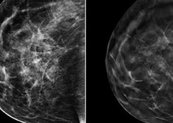
Breast density increases the risk of cancer and hinders mammographic detection [Classics Series]
This study summary is an excerpt from the book 2 Minute Medicine’s The Classics in Medicine: Summaries of the Landmark Trials
1. Women with mammographic density obscuring more than 75% of the imaged breast demonstrated a significantly increased risk of breast cancer detected either by screening or within 12 months of a negative screening examination, compared to those with density in less than 10% of the mammogram.
2. The increase in risk of breast cancer in women with dense breasts was greatest in younger women. In this demographic, 25% of all detected cancers were attributable to density in at least 50% of the mammogram.
Original Date of Publication: January 2007
Study Rundown: Mammography is an X-ray based modality, visualizing the various tissues of the breast based on their inherent attenuation properties. Fat appears radiolucent, while epithelial and stromal tissues appear radiodense. As the relative proportion between these tissues shifts away from fat, the breast becomes increasingly “dense” on mammography. As many tumors may also appear radiodense, they may be hidden by the predominance of stroma within a dense breast, subsequently hindering early detection, while that increased density may itself be a risk factor for oncogenesis. Unfortunately, this interaction between breast density and the ability to detect cancers on screening previously confounded research regarding the risks associated with mammographic density. Cancer risk would be underestimated if based solely on those tumors detected at screening due to an omission of those masked by breast density, while risk would be overestimated if based solely on cancers detected by alternate means, as it would focus only on those missed at screening. The present study attempted to better estimate the true risk of mammographic density as it relates to breast cancer by determining the risk both at the time of screening and thereafter among women with quantified breast density from three large national case-control studies. Women with density in 75% or more of the mammogram demonstrated a significantly increased risk of breast cancer compared to those with density in 10% or less of the mammogram both at the time of screening or by alternate means. This risk was found to persist for up to 8 years after initial screening. The attributable risk of cancer to breast density was most marked in younger women, for whom 26% of all detected cancers, and 50% of those detected within 1 year of a negative examination were attributable to density in at least 50% of the mammogram. This study established an accurate estimate of the incidence of breast cancers attributable to increased breast density by combining those data for those detected at screening and those detected up to 12 months after a negative screening examination, representing cancers likely present at the time of initial screening but obscured by mammographic density.
Click to read the study in NEJM
In-Depth [case-control study]: This retrospective case-control study pooled data from three large mammographic screening programs in Canada, the National Breast Screening Study (NBSS), the Ontario Breast Screening Program (OBSP), and the Screening Mammography Program of British Columbia (SMPBC.) A total of 1114 case-control pairs were included (mean age 56.7±9.1 years). Case subjects had histologically verified invasive breast cancer, excluding those with cancers detected within 12 months of their first screening mammogram, while up to 10 control subjects were matched to each index case on the basis of age, body-mass index, pregnancy and menopause history, hormone-replacement use, family history of breast cancer, and quantification of mammographic density. Mammograms from each subject were submitted and independently classified into one of six quantified degrees of breast density (0%, <10%, 10 to <25%, 25 to <50%, 50 to <75%, and ³75%) by two radiologists and a single observer using a computer-assisted technique. Notably, the average percentage of mammographic density was 5.8% higher among case subjects than control subjects at baseline. Women below the median age of 56 years were 3 times more likely to have breasts with a mammographic density of ³50%. The combined odds ratio for the detection of breast cancer among women with ³75% mammographic density was 4.7 (95%CI 3.0-7.4) as compared to those with £10% density. For those cancers detected at screening, the odds ratio was 3.5 (95%CI 2.0-6.2), while it was 17.8 (95%CI 4.8-65.9) for those cancers detected within 12 months of a negative screening mammogram. For cancers detected over a year after the last negative screening mammogram, the odds ratio was 5.7 (95%CI 2.1-15.5). These increased risks were significantly associated with percentage of breast density at all time periods, up to 8 years after entry into the study (p <0.001). The attributable risk of breast cancer for a mammographic density of ³50% was 16% for all cancers, much of which was limited to the 12 months following screening, suggesting that tumor masking was the principal mechanism of risk, rather than rapid tumor growth.
Boyd NF, Guo H, Marin LJ, Sun L, Stone J, Fishell E, et al. Mammographic Density and the Risk and Detection of Breast Cancer. The New England Journal of Medicine. 2007 Jan 18;356(3):227–36.
©2022 2 Minute Medicine, Inc. All rights reserved. No works may be reproduced without expressed written consent from 2 Minute Medicine, Inc. Inquire about licensing here. No article should be construed as medical advice and is not intended as such by the authors or by 2 Minute Medicine, Inc.
RelatedReports

![Radiofrequency catheter ablation effective as first-line therapy for atrial fibrillation [RAAFT-2 trial]](https://www.2minutemedicine.com/wp-content/uploads/2014/02/afib_-75x75.jpg)
