
![Radiofrequency catheter ablation effective as first-line therapy for atrial fibrillation [RAAFT-2 trial]](https://www.2minutemedicine.com/wp-content/uploads/2014/02/afib_-75x75.jpg)


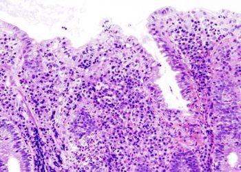
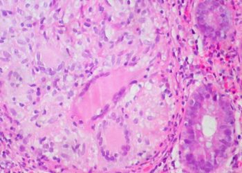
MRI offers an alternative to endoscopic assessment of disease activity and severity in ileocolonic Crohn’s disease [Classics Series]
This study summary is an excerpt from the book 2 Minute Medicine’s The Classics in Medicine: Summaries of the Landmark Trials
1. Magnetic resonance imaging (MRI) offers an effective alternative to endoscopy for quantifying disease severity in Crohn’s disease.
2. MRI required 3T field strength, both oral and rectal contrast, IV antiperistalsis medication, and IV contrast administration.
Original Date of Publication: January 2009
Study Rundown: Appropriate therapy of Crohn’s disease depends on disease severity and complications. The variable lifelong clinical course therefore necessitates frequent and accurate evaluation of disease activity. Historically, the diagnostic gold standard for Crohn’s disease is based on endoscopic findings coupled with biochemical markers (such as faecal calprotectin (fC) and C-reactive protein (CRP)) and clinical scores (Harvey-Bradshaw Index (HBI) and Crohn’s disease activity index (CDAI)). Endoscopy, however, only can access the proximal small bowel or terminal ileum. Imaging modalities have consequently been tested. Computed tomography (CT) enterography is limited use due to exposure to ionizing radiation. Transabdominal ultrasound lacks sensitivity and specificity and has high interobserver variability and is limited by overlying bowel gas. Magnetic resonance (MR) imaging lacks radiation exposure, has validated sensitivity and specificity, high soft-tissue resolution, and is therefore being increasing utilized in the diagnosis of Crohn’s disease. There was previously a lack of definitive criteria to evaluate Crohn’s disease based on MR parameters. In a 2008 cross-sectional study done at the Hospital Clinic de Barcelona, a Magnetic Resonance Index of Activity (MaRIA) using wall signal intensity (WSI), relative contrast enhancement (RCE), edema, and presence of ulcers was internally validated as independent predictors of Crohn’s disease severity. The same researchers externally validated MaRIA 3 years layer. Given the segmental predictors MaRIA uses, however, it may fail to appreciate the wide-ranging complications and total disease burden of Crohn’s disease. In a 2013 prospective trial by Makanyanga et al., a magnetic resonance enterography global score (MEGS) was internally validated against fC and CRP. Used in conjunction, the measures provide an accurate measure of the activity and severity of Crohn’s disease.
Click to read the study in Gut
In-Depth [cross-sectional]: This cross-sectional assessed 50 patients with clinically active (n=35) or inactive (n=15) Crohn’s disease based on the HBI. Ileocolonoscopy was used as the reference standard against MR. Except for a small number of patients, both assessments were performed the same day. Ileoendoscopic lesions were classified according to Crohn’s Disease Endoscopic Index of Severity (CDEIS). MR imaging was performed using a 3.0 T unit after bowel preparation and after oral ingestion of a large volume of polyethylene glycol. Additionally, saline was administered rectally and IV anti peristaltic medication was given. IV gadolinium was also administered. Bowel wall thickness, ulceration, edema, pseudopolyp identification, lymph node enlargement, WSI before and after contrast, and RCE were analyzed. The chi-square test, ANOVA followed by the Bonferoni posthoc test, and Spearman rank coefficient were used for statistical analysis; results were subsequently internally validated using bootstrap bagging with 1000 samples. All paired MR evaluations were performed by two radiologists for interobserver agreement. Complete endoscopic evaluation was completed in 36 patients (72% of total), yielding 213 colonic segments. MR and endoscopic findings were highly correlated for all parameters and interobserver agreement was very high (p <0.001 for all). Wall thickness (p <0.001), RCE (p = 0.013), and ulcer identification (p = 0.003) at MR were found to be independent predictors of active disease based on logistic regression analysis, with a sensitivity of 0.9 and specificity of 0.94. The MaRIA index was calculated to have a high (r = 0.81) and significant (p <0.001) correlation with the CDEIS of each analogous colonic segment. Overall, the index has a high accuracy for detecting Crohn’s disease activity (sensitivity 0.81, specificity 0.89) and for detecting ulcerative lesions (sensitivity 0.95, specificity 0.91) in the colon and terminal ileum.
Rimola J, Rodriguez S, García-Bosch O, Ordás I, Ayala E, Aceituno M, et al. Magnetic resonance for assessment of disease activity and severity in ileocolonic Crohn’s disease. Gut. 2009 Aug 1;58(8):1113–20.
Additional Review:
Makanyanga JC, Pendsé D, Dikaios N, Bloom S, McCartney S, Helbren E, et al. Evaluation of Crohn’s disease activity: Initial validation of a magnetic resonance enterography global score (MEGS) against faecal calprotectin. Eur Radiol. 2014 Feb 1;24(2):277–87.
Rimola J, Ordás I, Rodriguez S, García-Bosch O, Aceituno M, Llach J, et al. Magnetic resonance imaging for evaluation of Crohn’s disease: validation of parameters of severity and quantitative index of activity. Inflamm Bowel Dis. 2011 Aug;17(8):1759–68.
©2022 2 Minute Medicine, Inc. All rights reserved. No works may be reproduced without expressed written consent from 2 Minute Medicine, Inc. Inquire about licensing here. No article should be construed as medical advice and is not intended as such by the authors or by 2 Minute Medicine, Inc.
RelatedReports
