

Patient Basics: Computed Tomography (CT Scan) for Back Problems
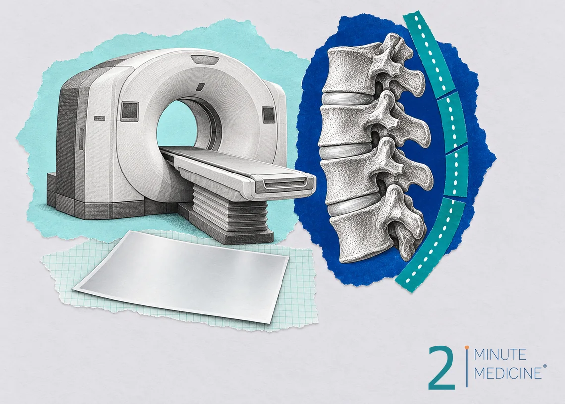
A back CT scan (computed tomography, sometimes called a CAT scan) uses X-rays and a computer to make detailed cross-section pictures of the bones of your spine. It is the best test for broken bones in the back, and it also shows arthritis, bone spurs and narrowing around the nerves. Most back pain gets better without any scan, so doctors save CT for injuries, warning signs or planning treatment. The scan itself takes only a few minutes.
Key points
- CT shows bone in fine detail. MRI is usually better for discs, nerves and the spinal cord.
- For new low back pain without warning signs, guidelines advise waiting about 6 weeks before any imaging, because most pain improves on its own.
- You lie on a table that slides through a large, open ring. It is quick and painless.
- A lower back CT uses more radiation than a plain X-ray, roughly as much as you’d get from natural sources in about 2 years.
- Disc bulges and wear-and-tear changes are common on scans of people with no pain, so results must be matched to your symptoms.
When a back CT scan is used
- After an injury, such as a car crash or a bad fall, to look for broken vertebrae
- Possible compression fractures from thinning bones (osteoporosis), especially in older adults with sudden back pain
- Spinal stenosis, a narrowing of the space around the nerves that can cause leg pain when walking
- Planning surgery, or checking how bones have healed or fused and where screws or rods sit after surgery
- When MRI isn’t possible, for example because of a pacemaker, certain metal implants or severe fear of closed spaces
- Guiding a needle for a biopsy or a pain injection
For infection, cancer, or a nerve pinched by a disc, MRI is usually the first choice. Sometimes CT is combined with dye placed in the spinal canal (a CT myelogram) to show the nerves when MRI can’t be done.
What a CT scan can and can’t show
CT clearly shows fractures, bone spurs, arthritis in the small joints of the spine, slipped vertebrae (spondylolisthesis), and narrowing of the spinal canal. It can show a herniated disc, though with less detail than MRI. It can’t show a pulled muscle, a strained ligament, or an irritated nerve, which cause much ordinary back pain.
Getting ready for a back CT scan
- Most back CT scans need no special preparation, and you can eat as usual.
- Wear clothes without metal, or change into a gown. Remove belts, jewelry and anything metal near your back.
- Tell the staff if you are or could be pregnant.
- Contrast dye is rarely needed for a back CT. If it is, tell the staff about kidney problems, diabetes medicines and any past reaction to X-ray dye.
- For a CT myelogram, you’ll get separate instructions about blood thinners and a ride home.
During the scan
You lie face up on a narrow table. The table slides slowly through the opening of a large, ring-shaped scanner. The ring is short and open on both ends, so most people don’t feel closed in. You’ll hear whirring and clicking. The technologist talks to you from the next room and may ask you to stop breathing for a moment. Lying still keeps the pictures sharp.
The scanning takes less than a minute or two, and the whole visit usually takes 15 to 30 minutes. If lying flat hurts, tell the technologist. Pillows under your knees can help.
Afterward
You can go back to your normal activities right away. If you had contrast dye, drink extra fluids that day. After a CT myelogram, you’ll rest with your head raised for a few hours, and you may get a headache.
Possible risks
- Radiation. The dose is small, but repeated scans add up. Ask whether a scan will change your treatment.
- Pregnancy. A back CT may expose the baby to radiation, so other tests are usually chosen.
- Contrast reactions, such as itching, hives or, rarely, breathing trouble, if dye is used.
- Findings that cause worry. Scans often show harmless changes that can lead to more tests or treatments you don’t need.
Your results
A radiologist reads the scan and sends a report to your doctor, usually within a day or two. Reports often use terms such as “degenerative disc disease,” “disc bulge,” “facet arthritis” or “foraminal narrowing.” These describe normal aging in many people. Your doctor will explain which findings, if any, explain your pain.
When to call your doctor
Most back pain improves with time, staying active and simple pain relief. See a doctor if pain lasts more than a few weeks, keeps you up at night, or spreads down a leg.
Get emergency care for back pain with:
- Numbness in the groin, buttocks or inner thighs
- New trouble controlling your bladder or bowels
- Weakness in one or both legs that is getting worse
- Fever, or a recent serious fall or crash
Common questions about back CT scans
Is a CT or an MRI better for back pain?
It depends on what your doctor is looking for. CT is faster and better for bones, so it’s the usual choice after an injury. MRI is better for discs, nerves, the spinal cord, infection and tumors, and it uses no radiation. For most long-lasting back pain with leg symptoms, MRI comes first.
Can a CT scan show a herniated disc?
Yes, often, especially in the lower back. A CT can show a disc pushing into the spinal canal. MRI shows discs and the nerves they press on in more detail, so doctors usually prefer it unless you can’t have one. A CT myelogram is another option in that case.
Why won’t my doctor order a scan for my back pain?
Most new back pain comes from muscles and joints and gets better within a few weeks without a scan. Early imaging rarely changes treatment, adds radiation, and often finds harmless changes that cause worry. Doctors order a scan when there are warning signs, after an injury, or when pain doesn’t improve.
Questions to ask your doctor
- What are you looking for with this scan?
- Would an MRI or an X-ray be better for me?
- Will the results change my treatment?
- Which findings on my report explain my symptoms?
- What can I do to ease my pain while I wait?
Learn more
- CT scans (MedlinePlus, U.S. National Library of Medicine)
- Back pain (National Institute of Arthritis and Musculoskeletal and Skin Diseases)
This guide is general information and doesn’t replace advice from your own doctor. Last updated October 2026.
RelatedReports







 Get the app