

Patient Basics: Echocardiogram
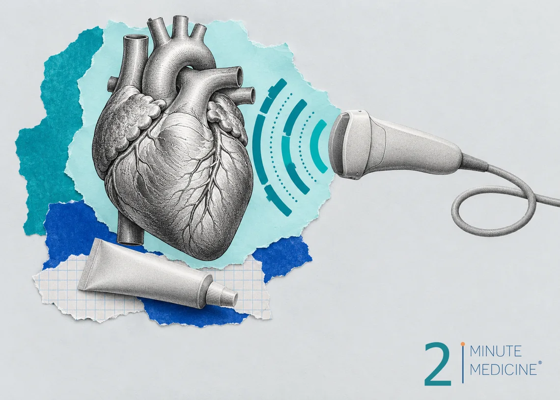
An echocardiogram, often called an echo, is an ultrasound test of the heart. It uses sound waves to make moving pictures of your heart’s chambers, valves and walls, so your doctor can see how well it pumps. The standard echo is painless, uses no radiation and needs no special preparation.
Key points
- The usual echo is done from the outside of the chest and takes about 30 to 60 minutes.
- It shows the heart’s size, pumping strength, valves and the sac around it.
- Some types need preparation. A transesophageal echo (TEE) means fasting and a ride home, and a stress echo means exercise or medicine.
- You can eat, drink and take your usual medicines before a standard echo.
- Results often come back within a few days.
Why an echocardiogram is done
Doctors order an echo to look for the cause of symptoms or to follow a known heart problem. Common reasons include:
- Shortness of breath, swollen legs or tiredness that could mean heart failure
- A heart murmur, an extra sound that may come from a leaky or narrowed valve
- Chest pain, fainting or an abnormal ECG (EKG)
- Checking the heart after a heart attack, or before and after heart surgery
- Looking for a blood clot or a small hole between the heart’s upper chambers after a stroke
- Checking the heart during some cancer treatments that can weaken the heart muscle
- High blood pressure in the lungs (pulmonary hypertension)
Types of echo test
- Transthoracic echo (TTE). The most common type. A probe moves over the skin of your chest.
- Transesophageal echo (TEE). A thin, flexible probe goes down your throat into the food pipe (esophagus), which sits right behind the heart. It gives clearer pictures of the valves and the upper chambers, and it is often used to look for clots or infection on a valve.
- Stress echo. Pictures are taken before and right after you walk on a treadmill or ride a bike, or while you get a medicine that makes the heart work harder. Areas that don’t move well under stress can point to narrowed heart arteries.
- Contrast or bubble study. A liquid injected into a vein makes the inside of the heart show up better, or shows whether tiny bubbles pass through a hole between chambers.
- Fetal echo. An ultrasound of an unborn baby’s heart, done through the mother’s belly during pregnancy.
How to prepare for an echocardiogram
- Standard echo: No fasting. Take your usual medicines. Wear a top that’s easy to remove, and skip lotion on your chest.
- TEE: Don’t eat or drink for about 6 hours before. Tell the team if you have trouble swallowing or a problem with your esophagus. You’ll get sedation, so arrange for someone to drive you home.
- Stress echo: Wear comfortable shoes and clothes for walking. You may be asked to skip caffeine and some heart medicines, such as beta-blockers, beforehand. Don’t stop any medicine unless your doctor tells you to.
During the test
You’ll undress from the waist up and put on a gown that opens in the front. You lie on your left side on an exam table, which brings the heart closer to the chest wall. Small sticky pads on your chest track your heart rhythm.
A trained technician (sonographer) spreads cool gel on your chest and presses a handheld probe against it at different spots, including below the ribs and at the base of the neck. You may hear a whooshing sound, which is blood flow. You might be asked to hold your breath or roll slightly. The pressure can feel firm but shouldn’t hurt.
For women, the sonographer may need to lift or move the left breast gently to reach the heart. You stay covered as much as possible, and you can ask for a female sonographer or a chaperone.
For a TEE, your throat is numbed with a spray and you get medicine through an IV to relax. The doctor guides the probe down as you swallow. It takes about 15 to 30 minutes, and most people remember little of it.
Afterward and possible risks
After a standard echo you wipe off the gel and go about your day. It has no known risks. A stress echo can bring on chest pain or an abnormal rhythm, which is why staff watch you closely. After a TEE you can’t eat until your throat is no longer numb, usually about an hour, and your throat may be sore for a day. Serious problems, such as a tear in the esophagus, are rare. Contrast agents can rarely cause an allergic reaction.
Your echocardiogram results
A heart specialist (cardiologist) reviews the pictures. Your report may mention:
- Ejection fraction (EF). The share of blood the main pumping chamber pushes out with each beat. Normal is roughly 50% to 70%. A lower number can mean a weak heart muscle.
- Valves. Whether any valve leaks (regurgitation) or is narrowed (stenosis), and how badly.
- Chamber size and wall thickness. Enlarged chambers or thick walls can come from high blood pressure, valve disease or problems with the heart muscle.
- Fluid around the heart (pericardial effusion), clots or holes.
Minor findings, such as a trace valve leak, are common in healthy people and often need no treatment.
When to call your doctor
- You haven’t heard about your results within a week
- Your throat hurts badly, you can’t swallow, or you have a fever after a TEE
Call 911 for chest pain, fainting or severe shortness of breath at any time.
Common questions about echocardiograms
Is an echocardiogram the same as an EKG?
No. An EKG (ECG) records the heart’s electrical signals with stickers on the skin and takes a few minutes. An echocardiogram uses ultrasound to show the heart’s structure and movement. They answer different questions, and doctors often order both.
Can an echo show blocked arteries?
Not directly. A standard echo can’t see the small arteries on the heart’s surface. It can show heart muscle damaged by an earlier heart attack. A stress echo can suggest narrowed arteries, and a CT scan or a cardiac catheterization shows the arteries themselves.
Can I eat before an echocardiogram?
Yes, for a standard echo you can eat, drink and take your usual medicines. The rules change for other types. Fast for about 6 hours before a TEE, and follow your clinic’s advice about caffeine and meals before a stress echo.
Is an echocardiogram safe during pregnancy?
Yes. A standard echo uses sound waves, not radiation, and is commonly done during pregnancy to check the mother’s heart. A fetal echo looks at the baby’s heart, usually around 18 to 24 weeks, when there is a reason to check it.
Questions to ask your doctor
- Which type of echo do I need, and why?
- Do I need to change my medicines or meals beforehand?
- What is my ejection fraction?
- Do my valves look normal?
- Will I need a repeat echo, and when?
Learn more
- Echocardiogram (MedlinePlus, U.S. National Library of Medicine)
This guide is general information and doesn’t replace advice from your own doctor. Last updated October 2026.
RelatedReports







 Get the app