
Transplantation of genetically altered stem cells improves bone formation in mice [PreClinical]
1. Hematopoietic stem cells (HSCs) were engineered to moderately overexpress platelet-derived growth factor (PDGF-BB), an important bone growth factor.
2. Mice transplanted with HSCs moderately overexpressing PDGF-BB showed increased levels of the bone formation marker alkaline phosphatase (ALP), bone formation, and bone strength compared to untreated mice or mice transplanted with HSCs minimally overexpressing PDGF-BB.
Evidence Rating Level: 3 (Average)
Study Rundown: Bone deterioration characteristic of osteoporosis is responsible for millions of fractures worldwide each year. While current therapies largely focus on drugs which prevent bone resorption or promote bone restoration, the authors of this study approached osteoporosis treatment using cell therapy. In this work, they transplanted mice with HSCs engineered to overexpress the protein PDGF-BB from its gene, PDGFB, with the goal of increasing bone density and strength.
Genetic promoters of varying strengths were used to induce PDGFB overexpression in HSCs. While use of a strong promoter led to new bone formation in mice, high PDGF-BB levels actually caused softening rather than strengthening of newly formed bone. Use of a more moderate promoter, phosphoglycerate kinase (PGK), resulted in similar serum PDGF-BB levels compared to controls or when a weaker P200 promoter was used. However, ALP levels were significantly higher in PGK-PDGFB mice than control mice and P200-PDGFB mice. Femurs from mice in the PGK-PDGFB group also showed a greater number of highly connected trabeculae and increased strength and stiffness as compared to femurs from control and P200-PDGFB mice. Subsequent experiments demonstrated that PGK-PDGFB HSC transplantation promoted bone marrow mesenchymal stem cell (MSC) formation, angiogenesis, and bone turnover.
Since this study was performed on mice with normal bone densities, it will be critical for the authors to test PGK-PDGFB HSC therapy in animal osteoporosis models before applying it to human medicine. Further tests should also include additional measures of bone strength after PGK-PDGFB HSC transplantation. PDGF-BB is already approved for use by the FDA for other clinical applications.
Click to read the study in PNAS
Relevant Reading: PDGF-BB secreted by preosteoclasts induces CD31hiEmcnhi vessel subtype in coupling osteogenesis
In-Depth [animal study]: HSCs were specifically chosen because they reside in the same sites where typical osteoporotic bone loss occurs. Mice transplanted with HSCs containing spleen focus-forming virus as a strong promoter to induce PDGF-BB overexpression showed newly formed bone in their femurs compared to controls at 4 weeks following transplantation. However, the bone was largely unmineralized, and higher serum PDGF-BB levels were associated with decreased trabecular bone mineral density (p<0.05, n=4-9).
When the more moderate PGK promoter was used, serum PDGF-BB levels did not differ from control or P200-PDGFB mice at 5 weeks following transplantation (n=8 per group). Serum ALP activity was higher in PGK-PDGFB mice than in control or P200-PDGFB mice (p<0.05, n=8 per group measured at 5-11 weeks following transplantation), and cytochemical staining showed increased ALP presence on the femoral surfaces of PGK-PDGFB mice compared to control mice (p<0.001, n=8 per group). At 12 weeks following transplantation, femoral trabecular bone volume as well as trabecular thickness, number, and connectivity density were all higher in PGK-PDGFB mice than control mice (p<0.001-0.01 at the femoral distal metaphysis and midshaft, n=6 per group). Importantly, femurs from PGK-PDGFB mice withstood a 45% heavier maximum load and were 46% stiffer than femurs from control or P200-PDGFB mice when subjected to three-point bending mechanical testing (p<0.01, n=6 per group).
Colony-forming unit (CFU) assays showed that PGK-PDGFB mice had more CFU-fibroblasts and CFU-osteoblasts, two types of MSCs, in their bone marrow compared to control mice (p<0.05, n not specified). Immunostaining showed greater angiogenesis in PGK-PDGFB mice, as well as increased presence of tartrate-resistant acid phosphatase, a marker of bone turnover (p<0.05, n=8 per group).
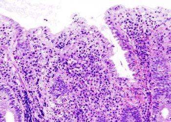
Image: CC/Wiki/Dilmen
©2015 2 Minute Medicine, Inc. All rights reserved. No works may be reproduced without expressed written consent from 2 Minute Medicine, Inc. Inquire about licensing here. No article should be construed as medical advice and is not intended as such by the authors or by 2 Minute Medicine, Inc.
RelatedReports


