
2 Minute Medicine Rewind December 18, 2017
A Bivalent Meningococcal B Vaccine in Adolescents and Young Adults
A vaccine against Neisseria meningitidis serotype B is needed, as current polysaccharide-based vaccines do not cover this serotype. A bivalent vaccine, MenB-FHbp, containing factor H-binding protein variants has been licensed in the US, however, large assessment of its coverage and protection against meningococcal disease has been limited. In this phase 3 randomized controlled trial, 3596 adolescents and 3304 young adults were randomized to receive either MenB-FHbp or saline at baseline, 2 months, and 6 months in order to study the effect on the generation of bactericidal antibodies in serum bactericidal assays with human complement (hSBAs). The five primary end points of the study were the proportion of participants who had an increase in their hSBA titer for each of the 4 primary strains by a factor of 4 or more, and the proportion of those who had an hSBA titer at least as high as the lower limit of quantitation for all 4 strains combined after dose 3. Researchers found that the percentages of vaccinated adolescents in whom the hSBA titers against four primary meningococcal B test strains increased by a factor of at least 4 ranged from 78.8% to 90.2% after dose 3. In vaccinated young adults, the percentage of participants in whom the hSBA titers increased by a factor of at least 4 from 78.9% to 89.7% after dose 3. Control participants who received saline showed negligible hSBA responses. In addition, an analysis of serum from participants showed that responses to the 4 primary strains were predictive of serum responses to 10 additional strains of meningococcus B. Overall, results from this trial support the use of the MenB-FHbp as a vaccine against meningococcal B disease. Further analysis is needed regarding the prevention of clinical disease, although this may be difficult due to the relatively low incidence of meningococcal B disease.
Noninvasive Cardiac Radiation for Ablation of Ventricular Tachycardia
Stereotactic body radiation therapy (SBRT) has been proposed as a noninvasive ablation approach that can be combined with noninvasive imaging to treat patients with refractory ventricular tachycardia. In this case series study, 5 patients with high-risk refractory ventricular tachycardia underwent SBRT with noninvasive electrocardiographic imaging in order to study the effect of treatment on the rate of ventricular tachycardia episodes. Before treatment, patients had an aggregate of 6577 episodes of ventricular tachycardia in the 15 patient-months before treatment. During the 6 weeks immediately after ablation, also known as the “blanking period”, where arrhythmias may occur due to post-ablation inflammation, there were 680 episodes of ventricular tachycardia. This was followed by a significant reduction in episodes of ventricular tachycardia during the next 46 patient-months (relative reduction of 99.9% from baseline), where only 4 episodes of ventricular tachycardia occurred. In terms of adverse effects, serial CT at 3 months showed inflammatory changes in the adjacent lung tissue that was typical of thoracic SBRT, however, there was near-complete resolution at 12 months. One patient had a fatal stroke 3 weeks after treatment, although it is unclear whether the stroke was associated with SBRT or preexisting medical conditions. Overall, results from this study suggest that the use of noninvasive stereotactic cardiac radiotherapy as an ablation technique can cause a marked reduction in the burden of ventricular tachycardia after treatment. Additional studies are needed in evaluating the safety and efficacy of SBRT and to study potential late effects of high-dose SBRT.
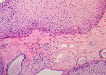
In pathology, computer-aided diagnostics that employ deep learning algorithms have been proposed as a way of improving diagnostic accuracy and efficiency. However, the discriminative accuracy of deep learning algorithms has not been well explored. In the Netherlands, a research challenge competition (CAMELYON16) was carried out to develop automated solutions for detecting lymph node metastases. As part of the competition, participating researchers were provided with a training data set of whole-slide images from 2 centers in the Netherlands with (n=110) and without (n =160) nodal metastases verified by immunohistochemical staining. Algorithm performance was evaluated in an independent test set of 129 whole-slide images (49 with and 80 without metastases). The same test set of corresponding glass slides was also evaluated by a panel of 11 pathologists with a time constraint to determine the likelihood of nodal metastases for each slide in a flexible 2-hour session, simulating routine pathology workflow, and by 1 pathologist without time constraint. The algorithms and pathologists were compared for two tasks: identification of individual metastases in whole-slide images (task 1) and classification of every whole-slide image as either containing or lacking SLN metastases (task 2). Results showed that out of the 32 algorithms developed, the best algorithm for task 1 achieved an overall true-positive fraction score of 80.7% (95% CI 73.2% to 88.9%), which was comparable to the true-positive fraction score of 72.4% (95% CI 64.3% to 80.4%) achieved by pathologists without time constraint. For task 2, the best algorithm had an area under the curve (AUC) of 0.994 (95% CI 0.938 to 0.999). This AUC was comparable to the AUC of 0.966 (95% CI 0.927 to 0.998) achieved by pathologists without time constraint, and was significantly higher than the mean AUC of 0.810 (range 0.738 to 0.884) achieved by pathologists with time constraint (p<0.001). Overall, 7 of the 32 submitted algorithms had a significantly higher AUC than the pathologists with time constraints (range of p<0.001 to p<0.04). Taken together, the results from this study indicate that deep learning algorithms can achieve diagnostic performance on whole-slide images that is comparable or better than the performance of pathologists working under time constraint. It is important to note, however, that conditions of the study are not necessarily reflective of what is encountered in clinical practice, and the algorithm runtimes were not recorded. As such, these algorithms need further assessment in a clinical setting.
High-sensitivity troponin (hsTnT) assays have been proposed as a way of identifying patients at low risk of experiencing 30-day adverse cardiac events (ACE). Identifying these patients is critical in avoiding the risk of unnecessary hospitalization but also the harms associated with inappropriate discharge from the emergency department (ED). In this prospective cohort study, investigators collected serial blood samples from 1355 individuals that presented to the ED with suspected acute coronary syndrome in order to correlate hsTnT measurements to the 30-day ACE rate. Investigators also collected blood samples from 1312 healthy individuals to establish a reference range. Researchers found that the 99% percentile upper reference level (URL) for healthy individuals was a hsTnT concentration of 19 ng/L. Patients in the ED who had a hsTnT level exceeding the URL had a 30-day ACE rate of 2.8%, whereas patients with a level below the URL had a rate of 0.7%. Serial hsTnT levels less than the URL of 19 ng/L resulted in a 99.3% negative predictive value for 30-day ACE (95% CI 99.1% to 99.6%). Using a cutoff of less than 6 ng/L for baseline hsTnT concentrations provided a negative predictive value of 99.4% (95% CI 98.6% to 99.8%) for acute myocardial infarction, and at 3 hours a less than 6 ng/L cutoff gave a negative predictive value of 99.7% (95% CI 99.0 to 100.0). Overall, results from this study indicate that serial hsTnT measurements can identify patients with 30-day ACE rates less than 1% with high negative predictive values. It is important to note, however, that this study was an observational analysis and since clinicians were blinded to hsTnT measurements, and some patients with low hsTnT values were still hospitalized, which may have impacted outcome. Patients who potentially had elevations of hsTnT from recent cardiac events or renal insufficiency were also not included in this study.
The increased use of prescription opioids in the US is the result of a multitude of factors, including changing attitudes towards pain relief. The increasing use of opioids, however, may also be attributed, in part, to drug diversion within households. In this retrospective cohort study, investigators compared 12,695,280 commercial insurance beneficiaries with a household member who started a new prescription of opioids to 6,359,639 beneficiaries with a household member who started a new prescription of non-opioid NSAID pain relievers in order to examine the effect of having a household member on prescription opioids on the 1-year risk of subsequent opioid use. To this end, investigators used data from the 2000-2014 Truven Health Analytics MarketScan Commercial Claims and Encounters databases. Inverse probability-weighted analysis of the data showed that the 1-year risk of prescription opioid initiation was 11.83% (95% CI 11.81% to 11.85%) for opioid households and 11.11% (95% CI 11.09% to 11.14%) for NSAID households, yielding an adjusted 1-year risk difference in opioid initiation of 0.71% (95% CI 0.68% to 0.74%). Overall, results from this study suggest that living in a household with a prescription opioid user may increase the risk of prescription opioid use; this result has implications for what considerations need to be made in initiating opioid prescriptions, taking into account factors such as increased access and shared risk factors. This study was limited, however, as it used electronic pharmacy dispensing billing claims to analyze opioid prescription and therefore did not include opioids paid for in cash or those received in inpatient settings. The MarketScan databases also oversamples the South and has limited coverage of the West, and so results from this study may not be directly generalizable to the US population as a whole.
Image: PD
©2017 2 Minute Medicine, Inc. All rights reserved. No works may be reproduced without expressed written consent from 2 Minute Medicine, Inc. Inquire about licensing here. No article should be construed as medical advice and is not intended as such by the authors or by 2 Minute Medicine, Inc.
RelatedReports


