
Diffusion-weighted imaging may aid in prognostication for intracerebral hemorrhage
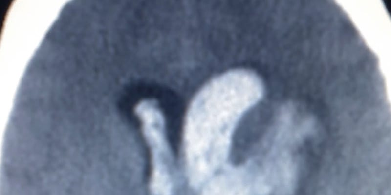
1. Diffusion-weighted imaging (DWI)-positive lesions are common after intracerebral hemorrhage (ICH) and serve as an independent biomarker of cerebral small vessel disease (cSVD) severity and worse 6-month functional outcome, supporting their potential use in prognostication and clinical trial patient selection.
2. The anatomical distribution of DWI-positive lesions may help distinguish cerebral amyloid angiopathy (CAA) from non-CAA small vessel arteriopathy, as deep lesions were never observed in probable CAA cases.
Evidence Rating Level: 2 (Good)
Diffusion-weighted imaging (DWI)-positive lesions are increasingly recognized in acute intracerebral hemorrhage (ICH) as markers of cerebral small vessel disease (cSVD), but their mechanisms and prognostic significance remain incompletely defined. This prospective cohort study analyzed 342 consecutive patients with acute ICH from the SIGNAL registry (North Central London, 2017–2020) who underwent diagnostic MRI, assessing DWI-positive lesions and STRIVE-2–defined cSVD markers, with the primary outcome being 6-month modified Rankin Scale (mRS) score. DWI-positive lesions were present in 21.6% of patients, most often in lobar regions. They were independently associated with severe white matter hyperintensities (OR 3.37), ICH volume ≥30 mL (OR 2.29), higher cerebral microbleed burden (OR 1.61), and poor 6-month functional outcome (OR 2.16). Patients with cerebral amyloid angiopathy (CAA) more frequently had strictly lobar DWI-positive lesions, whereas strictly deep lesions occurred exclusively in non-CAA patients, suggesting distribution reflects underlying arteriopathy.
Click here to read this study in Neurology
Image: PD
©2026 2 Minute Medicine, Inc. All rights reserved. No works may be reproduced without expressed written consent from 2 Minute Medicine, Inc. Inquire about licensing here. No article should be construed as medical advice and is not intended as such by the authors or by 2 Minute Medicine, Inc.