MRI screening may improve osteonecrosis outcomes in children with leukemia
1. In a prospective cohort of over 400 children with acute lymphoblastic leukemia treated with chemotherapy, hip MRI have high sensitivity and specificity for detecting glucocorticoid-induced osteonecrosis.
2. Use of screening in children older than 10 years of age within 1 year after initiation of glucocorticoids may delay and prevent future hip joint destruction and collapse.
Evidence Rating Level: 2 (Good)
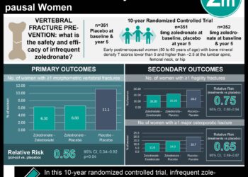
Study Rundown: Glucocorticoid-induced osteonecrosis of the hip is a common complication in children treated for acute lymphoblastic leukemia (ALL) and may lead to femoral head collapse and long-lasting disability. However, there is little data on the specific time of onset or the risk factors that increase the risk of osteonecrosis in this population. The purpose of this study was to evaluate the utility of early screening magnetic resonance imaging (MRI) in identifying asymptomatic osteonecrosis in children treated for ALL. The authors prospectively followed over 400 patients that underwent screening MRI of the hip at 6.5 months and 9 months into chemotherapy (early screen) and after (final screening) chemotherapy. At the conclusion of the study, the authors found that 22% of all ALL patients screened develop radiographic signs of osteonecrosis, with the majority of patients detected by 1 year after starting chemotherapy. Furthermore, age was associated with the severity of osteonecrosis and response to treatment. Age over 10 years was associated with more extensive osteonecrosis and increased rate of total hip arthroplasties compared to patients less than 10 years of age. The results of this study support the use of early screening for patients 10 years or older to prevent progressive joint damage. This study was strengthened by the size of the population screened and the defined glucocorticoid schedules within this cohort. The study was limited by relatively short follow-up. Additional longitudinal studies are needed to accurately describe the long-term outcomes of these patients.
Click to read the study in JCO
In-Depth [prospective cohort]: A cohort of 462 pediatric patients diagnosed with ALL and treated with the Total Therapy Study SV chemotherapy protocol from 2000-2007 were included in this study. Hip MRI was performed at weeks 10 and 20 of treatment, approximately 6.5 and 9 months from diagnosis (early screen). Patients were scanned again at completion of therapy (final screen). Patients were surveyed on symptoms of osteonecrosis during weekly clinic visits for chemotherapy. At the completion of the trial, the early screen and final screen detected osteonecrosis of at least one hip in 17.1% and 21.7% of patients, respectively. Patients greater than 10 years of age had significantly greater risk of extensive osteonecrosis compared to patients 10 years or younger (HR: 15.6; 95% CI: 6.7 to 36.2; p<0.001). In the 10 years or older group, MRI screening detected 40 hips with severe osteonecrosis, with 19 hips requiring total hip arthroplasty. No hips required arthroplasty in the 10 years or younger cohort. Overall, hip MRI screening sensitivity was 84.1% and specificity was 99.4%.
More from this author: Rituximab linked with reduced chronic immune disease following stem cell transplantation, High-dose prophylaxis for hemophilia increases costs with minimal benefit, Ambrisentan found ineffective against idiopathic pulmonary fibrosis
Image: PD
©2015 2 Minute Medicine, Inc. All rights reserved. No works may be reproduced without expressed written consent from 2 Minute Medicine, Inc. No article should be construed as medical advice and is not intended as such by the authors, editors, staff or by 2 Minute Medicine, Inc.
RelatedReports