
Rapid CT contrast washout differentiates adrenal adenomas from nonadenomas [Classics Series]
This study summary is an excerpt from the book 2 Minute Medicine’s The Classics in Medicine: Summaries of the Landmark Trials
1. The mean percentage of computed tomography (CT) contrast washout was significantly greater for adenomas compared to nonadenomas in all delayed scans.
2. Delayed enhanced CT scans discriminated adenomas from nonadenomas with high sensitivity and specificity at various time points, as early as 5 to 15 minutes after enhancement.
Original Date of Publication: March 1998
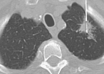
Study Rundown: Adrenal mass lesions are incidentally discovered in up to 5% of patients subjected to CT for indications unrelated to adrenal disease. In patients without a known history of cancer, most of these lesions are benign. For patients with a diagnosed extra-adrenal malignancy, the likelihood that an incidentally discovered adrenal lesion is malignant increases considerably. On unenhanced CT scans, benign adrenal adenomas are characterized by low attenuation. On contrast-enhanced CT, however, attenuation values cannot differentiate between benign and malignant lesions. As such, other imaging modalities or repeat imaging have been historically used to differentiate between benign and malignant adrenal masses. This was until several studies noted that intravenous (IV) contrast medium tended to “wash out” faster from adenomatous lesions than non-adenomatous lesions, and therefore, CT attenuation measurements could be used after a variable delay period to characterize adrenal lesions. Given that delayed CT attenuation measurements are dependent on the type, total dose and injection rate of IV contrast material, however, measures of absolute attenuation on delayed scans were found not found to be useful. In response, the use of enhancement washout curves was proposed to differentiate adrenal adenomas from nonadenomas. In this study conducted by Korobkin and colleagues, contrast enhancement washout curves were generated following delayed contrast-enhanced CT scans of adrenal adenomas and nonadenomas. The results of this study showed that the mean percentage of enhancement washout for adrenal adenomas far exceeded that observed in nonadenomas. The authors also demonstrated that delayed enhanced CT scans could discriminate adenomas from nonadenomas with high sensitivity and specificity at various time points, as early as 5 to 15 minutes after enhancement. Pena and colleagues later confirmed in their study that the relative percentage washout on dynamic and delayed enhanced CT scans could be used to characterize adrenal masses.
Click to read the study in ARRS
In-Depth [prospective cohort]: Patients with adrenal masses identified through abdominal or chest CT were consecutively enrolled into this study. Adrenal adenoma diagnoses were confirmed through various means, including percutaneous biopsy, stable appearance on follow-up CT examinations and an attenuation value of <10 Hounsfield Units (HU) on unenhanced CT. Nonadenoma diagnoses were confirmed through surgery, percutaneous biopsy, substantial growth or shrinkage at short-term follow-up, and stable CT for 1 case of myelolipoma. Unenhanced and standard enhanced scans were obtained for all adrenal masses, and delayed enhanced CT values were studied in 2 groups of patients. In the first group, delayed scans were obtained at 15, 30 and 45 minutes following the initial enhanced CT. In the second group, delayed scans were obtained at 5, 10 and 15 minutes after the initial enhanced CT. From these scans, percentages of initial enhancement at these time points were calculated and used to generate washout curves for adrenal adenomas and nonadenomas. Calculations of sensitivity and specificity for the diagnosis of adenoma using delayed enhanced CT were made after selecting an optimal threshold value. A total of 66 patients with 76 adrenal masses were evaluated. The masses consisted of 52 adenomas (n=45; mean age 64 years, range 43-80 years; 53% men) and 24 nonadenomas (n=21; mean age 60 years, range 31-76 years; 71% men). Consistent with previous studies, there was no significant difference in mean CT attenuation at initial enhancement for adenomatous versus nonadenomatous masses. However, a statistically significant difference in mean CT attenuation between adenomas and nonadenomas was observed for unenhanced (p < 0.001) and all delayed enhanced (p <0.001) scans. For unenhanced scans, an optimal threshold of 10 HU corresponded to a sensitivity of 87% and specificity of 100%. For the 15-minute delayed enhanced scan, the sensitivity and specificity were both 96% at a threshold of 37 HU. Differences in mean percentages of initial enhancement were also found to be statistically significant between the adenoma and nonadenoma groups at all delayed times (p < 0.001). The mean percentage of enhancement washout for adrenal adenomas was 51% at 5 minutes and 70% at 15 minutes, compared to 8% and 20% for nonadenomas, respectively. On the 15-minute delayed enhanced scans, an optimal threshold for adenoma diagnosis was established at 60% contrast enhancement washout, associated with a sensitivity of 88% and specificity of 96%.
Korobkin M, Brodeur FJ, Francis IR, Quint LE, Dunnick NR, Londy F. CT time-attenuation washout curves of adrenal adenomas and nonadenomas. American Journal of Roentgenology. 1998 Mar;170(3):747-52.
Additional Review:
Blake MA, Cronin CG, Boland GW. Adrenal imaging. American Journal of Roentgenology. 2010;194(6):1450.
Boland GWL, Blake MA, Hahn PF, Mayo-Smith WW. Incidental Adrenal Lesions: Principles, Techniques, and Algorithms for Imaging Characterization. Radiology. 2008 Dec;249(3):756-75.
Peña CS, Boland GWL, Hahn PF, Lee MJ, Mueller PR. Characterization of Indeterminate (Lipid-poor) Adrenal Masses: Use of Washout Characteristics at Contrast-enhanced CT. Radiology. 2000 Dec;217(3):798-802.
©2022 2 Minute Medicine, Inc. All rights reserved. No works may be reproduced without expressed written consent from 2 Minute Medicine, Inc. Inquire about licensing here. No article should be construed as medical advice and is not intended as such by the authors or by 2 Minute Medicine, Inc.
![2MM: AI Roundup- AI Cancer Test, Smarter Hospitals, Faster Drug Discovery, and Mental Health Tech [May 2nd, 2025]](https://www.2minutemedicine.com/wp-content/uploads/2025/05/Untitled-design-350x250.png)

