

Canadian C-Spine Rules superior to NEXUS Low-Risk Criteria for detecting cervical-spine injuries [Classics Series]
![ABCD2 Score: Predicting Early Stroke Risk After Transient Ischemic Attack (TIA) [Classics Series]](https://www.2minutemedicine.com/wp-content/uploads/2013/05/web-cover-classics-with-logo-medicine-BW-small-jpg.jpg)
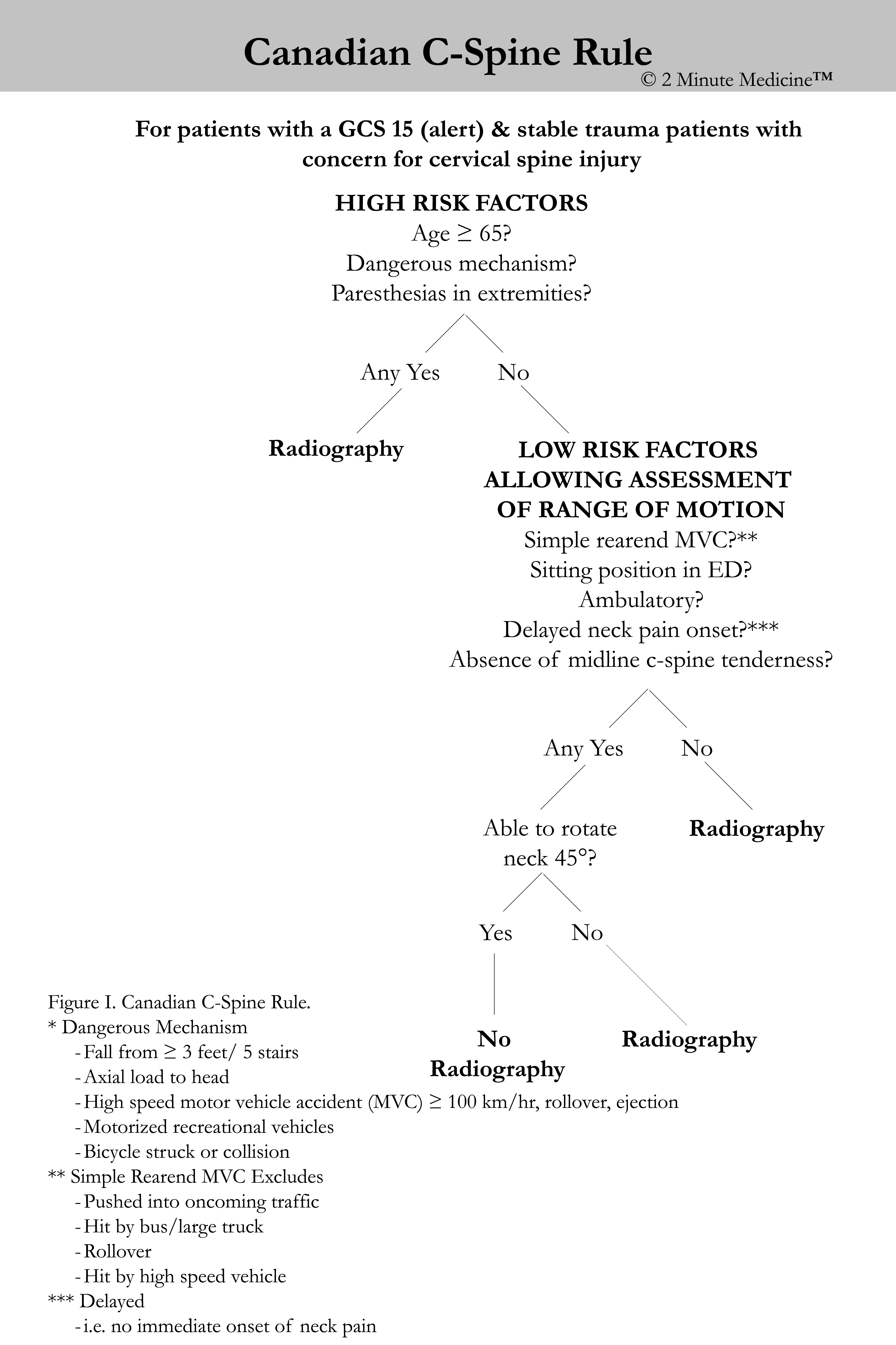
1. The Canadian C-Spine Rule (CCR, Figure I) demonstrated higher sensitivity and specificity for cervical-spine injury compared to the National Emergency X-Radiography Utilization Study (NEXUS) Low-Risk Criteria (NLC, Table I).
Original Date of Publication: December 2003
This study summary is an excerpt from the book 2 Minute Medicine’s The Classics in Medicine: Summaries of the Landmark Trials, 2e (The Classics Series).
Study Rundown: According to large trauma registries, approximately 3% of all blunt trauma victims sustain injuries to the spinal column, including injuries to the cervical spine (C-spine). Suspected C-spine fractures are frequently evaluated with radiography; however, there is a lack of consensus among published guidelines with regards to the use of radiography. Two independently validated clinical decision rules are commonly used to determine patient selection of C-spine radiography: the CCR and the NLC. The CCR is a three-step process that evaluates risk factors for serious injury, physical exam findings, and the ability for the patient to rotate their neck. The NLC is made up of five criteria based on patient history (no painful distracting injuries, normal level of consciousness) and physical examination (no midline cervical tenderness, no focal neurologic deficit, and no evidence of intoxication). The purpose of this landmark prospective trial was to compare the accuracy of both the CCR and the NLC in detecting C-spine injuries.
The prospective trial measured the frequency of clinically important C-spine injuries in over 8000 consecutive patients and reported the accuracy of both the CCR and NLC in detecting these injuries when applied to this cohort. At the conclusion of the trial, the CCR demonstrated a near 100% sensitivity for detecting clinically important C-spine fractures; this was significantly higher than the NLC (90.7%). Additionally, the CCR was also found to be more specific for C-spine injuries compared to NLC. When the sensitivity and specificity were applied to the cohort, the NLC would have missed 1 in 10 clinically important C-spine fractures while requiring a higher radiography rate. Ten percent (10%) of patients were not evaluated for neck range of motion and therefore did not complete the CCR algorithm. However, the sensitivity and specificity of the CCR remained significantly higher than the NLC even if all significant findings in this small subgroup were assumed to be missed by CCR.
The results of this trial demonstrated superior performance of the CCR over the NLC in detecting clinically significant C-spine injuries and supported the use of CCR as the preferred clinical prediction rule. However, the study population was only limited to patients aged greater than 16 years who were alert and in stable condition at the time of assessment. Additionally, the study was performed at institutions where the CCR was developed, precluding validation in alternative clinical settings.
Please click to read study in NEJM
In-Depth [prospective cohort]: This was a prospective study of 8282 consecutive patients with acute trauma to the head and neck for assessment for C-spine injuries in nine tertiary care hospitals in Canada. Patients were included if they were at least 16 years of age and had a Glasgow Coma Scale score of 15 out of 15 at the time of assessment. Patients were excluded if they had penetrating neck trauma, acute paralysis, or were pregnant. All patients were assessed by the CCR and NLC for the study, which was completed by the evaluating emergency physician. Patients underwent plain radiography according to the judgement of the physician independent of the results of the clinical prediction rules. All injuries to the C-spine on radiography were considered significant unless one of the following benign fractures were isolated: transverse process not involving a facet joint, a spinous process not involving lamina, or simple vertebral compression of < 25% of body height. Patients who did not receive radiography were evaluated for C-spine injuries using the Proxy Outcome Assessment Tool, which assessed clinical symptoms 14-days following injury.
At the conclusion of the trial, a total of 169 patients (2%) demonstrated a clinically significant C-spine fracture. The sensitivity for detection of these C-spine fractures was significantly higher in the CCR compared to the NLC (99.4% versus 90.7%; p < 0.001). Additionally, the CCR also demonstrated significantly higher specificity for C-spine injuries compared to NLC (40.4% versus 33.0%; p < 0.001). In 845 patients (10.2%), the CCR assessment was indeterminate due to the lack of evaluating of neck range of motion. In secondary analysis, if all indeterminate cases were assumed to be negative by the CCR, the sensitivity and specificity was modified to 95.3% and 50.7%, respectively. This remained significantly higher when compared to the NLC.
Table I. NEXUS Low-Risk Criteria.
| NEXUS Low-Risk Criteria |
|---|
| Cervical-spine radiography is indicated for trauma unless the patient exhibits all of the following criteria: 1. No posterior midline cervical tenderness 2. No focal neurological deficit 3. Normal alertness 4. No evidence of intoxication 5. No painful distracting injury |
Stiell IG, Clement CM, McKnight RD, Brison R, Schull MJ, Rowe BH, et al. The Canadian C-Spine Rule versus the NEXUS Low-Risk Criteria in Patients with Trauma. The New England Journal of Medicine. 2003 Dec 25;349(26):2510–8.
© 2 Minute Medicine, Inc. All rights reserved. No works may be reproduced without expressed written consent from 2 Minute Medicine, Inc. Inquire about licensing here. No article should be construed as medical advice and is not intended as such by the authors or by 2 Minute Medicine, Inc.




![PFO closure equivalent to medical management in prevention of recurrent stroke [PC and RESPECT trials]](https://www.2minutemedicine.com/wp-content/uploads/2013/03/Echokardiogram_von_Atriumseptumdefekt_Ostium_secundum1-75x75.jpg)


