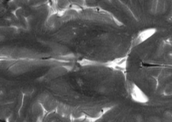
Diffusion tensor imaging may aid in Parkinson’s diagnosis
1. Diffusion tensor imaging (DTI) of marmoset brains following chemically-induced Parkinsonism showed an increase in nigrostriatial diffusivity with a loss of corresponding fiber tracts as compared to controls, indicating a potential future role for imaging in the diagnosis of Parkinson’s disease.
Evidence Rating Level: 3 (Average)
Study Rundown: In Parkinson’s disease (PD), degeneration of the nigrostriatal fibers projecting from the substantia nigra to the striatum are progressively lost, leading to the disease’s hallmark tremor and rigidity. This finding has been well defined histologically but is currently of limited use in the diagnosis and assessment of disease progression in a living patient. Diffusion tensor imaging (DTI) is a magnetic resonance-based imaging modality that allows for non-invasive visualization of fiber tracts in tissue based on the direction-dependent—or anisotropic—nature of water diffusion along such tracts. In the current study, a DTI-based tractographic examination of the difference in diffusivity and subsequent loss of fiber tracts along the nigrostriatal pathway was undertaken between normal marmosets and those treated with MPTP, a toxin specific to the dopaminergic neurons of the substantia nigra. In this chemically-induced primate model of Parkinsonism, the study authors found that DTI could be successfully used in detecting the nigrostriatal degeneration characteristic of PD, as represented by increase in localized axial diffusivity with a relative loss of fiber tracts. These findings were confirmed by subsequent histologic examination. The study was technologically limited by necessity for higher resolution imaging; particularly, it utilized long scan times and high field strengths, the use of which are currently limited to research use only. Future studies utilizing practical tractographic imaging parameters in humans with and without PD are warranted, as are additional primate studies examining a genetic, rather than chemically-induced, PD model.
Click to read the study in Radiology
Relevant Reading: Mapping track density changes in nigrostriatal and extranigal pathways in Parkinson’s disease
In-Depth [animal study]: Nine male marmosets underwent longitudinal, region-of-interest (ROI)-based DTI evaluation of the nigrostriatal pathway before and 10 weeks after MPTP administration using a 7T MR imaging system. Echo-planar DTI images were acquired continuously over the course of one week while animals were anesthetized and immobilized to reduce artifact degradation. The animals were subsequently euthanized for histologic and microscopic tractographic examination of the nigrostriatal pathway to confirm differences seen on longitudinal DTI. After MPTP administration, all animals displayed the characteristic symptoms of Parkinson disease, and a significant increase in the mean axial and radial diffusivity was seen bilaterally in the basal ganglia, extending up from the substantia nigra to the cerebral peduncles (P = 0.033). Specific ROI analysis of the nigrostriatal pathway revealed a 27% increase in diffusivity between the control and MPTP-treated states (P = 0.005 and P = 0.004 in the right and left cortices, respectively.) These findings were confirmed on histologic and microscopic tractographic examination, showing specific denervation of the nigrostriatal pathway and a significant reduction in fiber bundles in the MPTP-treated state.
More from this author: Perfusion imaging may predict successful thrombolysis in acute stroke, Stroke expansion following intra-arterial therapy may explain worse outcomes, Imaging of atherosclerotic inflammation possible with fluorine MRI
Image: CC/Jensflorian/Wikimedia Commons
©2014 2 Minute Medicine, Inc. All rights reserved. No works may be reproduced without expressed written consent from 2 Minute Medicine, Inc. No article should be construed as medical advice and is not intended as such by the authors, editors, staff or by 2 Minute Medicine, Inc.


