
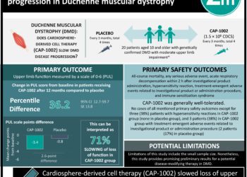
Gene therapy targets Duchenne muscular dystrophy in new trial [PreClinical]
1. Mutations in the gene encoding dystrophin, a large scaffolding protein that supports muscle cell structure, result in Duchenne muscular dystrophy (DMD).
2. A gene editing strategy to correct the dystrophin reading frame in a canine model of DMD is shown to partially restore dystrophin protein expression in skeletal and cardiac muscle and improve muscle histology.
Evidence Rating Level: 2 [Good]
Study Rundown: DMD leads to progressive muscle degeneration and premature death due to cardiac and respiratory muscle failure and occurs as a result of mutations in the dystrophin gene, which encodes a large scaffolding protein that helps link the muscle cytoskeleton to the plasma membrane. The dystrophin gene is large containing 79 exons. Many DMD-causing mutations occur in exons 45-50 and place exon 51 out of frame. Therapies that induce skipping of exon 51 thus restoring the open reading frame of the DMD gene can benefit DMD patients. This study aimed to demonstrate the safety and efficacy of a gene editing strategy for DMD in dogs, an essential step towards clinical translation in humans.
The deltaE50-MD canine model of DMD results in loss of exon 50, and can be corrected by skipping of exon 51. To correct the dystrophin reading frame in the deltaE50-MD canine model, a Cas9 enzyme coupled with a single guide RNA targeting the exon 51 splice acceptor site (sgRNA-51) was employed. Recombinant adeno-associated virus serotype 9 (AAV9), which has preferential tropism for skeletal and cardiac tissues, was used to deliver the Cas9 enzyme and targeting sgRNA into the cranial tibialis muscle of dogs. Deep sequencing revealed that ~10% of reads contained changes at the targeted genomic site, and further protein studies confirmed restoration of dystrophic expression to ~60% of wild-type levels. Systemic administration of viruses, in conjunction with high-dose immunosuppression, also resulted in partial restoration of dystrophin expression in heart and skeletal muscle without pronounced systemic side effects.
While this study holds promise for DMD gene therapy in humans, there still remain several safety concerns surrounding viral-vector based delivery systems for CRISPR-based gene editing. These concerns include potential off-target effects of in vivo gene editing, the immunogenicity of virus and Cas9 components, and the efficiency and heterogeneity of gene editing in target tissues.
Click here to read about animal models of Duchenne muscular dystrophy
Relevant Reading: Gene therapy for muscular dystrophy
In-Depth [animal study]: The objective of this study was to determine the therapeutic potential of in vivo CRISPR/Cas9 gene editing for the treatment of DMD in the ∆Ex50 dog model which has a naturally-occurring missense mutation in the 5’ donor splice site of exon 50 that results in deletion of exon 50. Short guide RNAs (sgRNAs) targeted Dmd exon 51 and were tested in tissue culture in primary myoblasts and MDCK cells prior to cloning into the rAAV9 backbone for in vivo administration. sgRNAs were placed under the transcriptional control of three different promoters (U6, H1, or 7SK), while Cas9 was placed under the control of a muscle-specific creatine kinase (CK) regulatory cassette. AAV9-Cas9 and AAV9-sgRNA were delivered to ∆Ex50 dogs via intramuscular injection in the left cranial tibialis muscle (n=2). In a separate experiment, viruses were administered systemically by delivery via the cephalic vein over 30 minutes in conjunction with a short-term prednisolone immunosuppressive regimen (n=2). For intramuscular injections, muscles were analyzed at 6 weeks post-injection, while for systemic administrations, muscles were analyzed at 8 weeks post-injection. Deep sequencing of target tissue revealed that ~10% of reads contained changes at the targeted genomic site including insertions, deletions, and substitutions. Reverse transcription-polymerase chain reaction (RT-PCR) analyses of the targeted tissues revealed that disruption of the exon 51 splice site resulted in deletion of exon 51, which allowed splicing from exon 49 to exon 52, restoring the dystrophin open reading frame. Western blot analyses confirmed that dystrophin expression was restored in treated skeletal muscle to ~60% of wild-type levels, albeit to varying levels throughout the tissue. Some dystrophin expression (~2% of wild-type level) was also detected in the uninjected contralateral muscles, suggesting some leakage of AAV9 into circulation. Systemic administration of viruses, in conjunction with a high-dose transient immunosuppressive regimen, also resulted in restored dystrophin expression in peripheral skeletal muscles, and to a lower extent in heart samples without any pronounced systemic side effects. The hematological and biochemical parameters of blood taken from the treated dogs compared to controls were unremarkable.
Image: PD/CDC
©2018 2 Minute Medicine, Inc. All rights reserved. No works may be reproduced without expressed written consent from 2 Minute Medicine, Inc. Inquire about licensing here. No article should be construed as medical advice and is not intended as such by the authors or by 2 Minute Medicine, Inc.


