

Patient Basics: Retrobulbar Neuritis (Optic Neuritis)
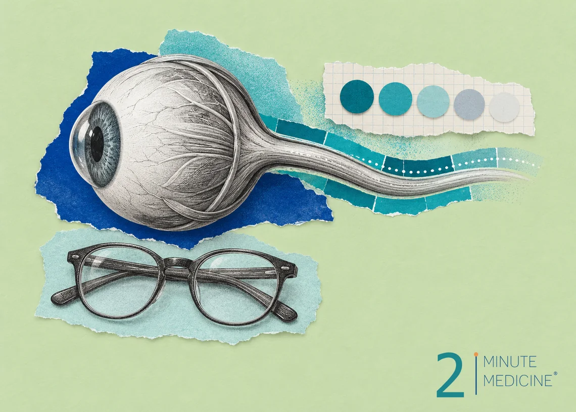
Retrobulbar neuritis is swelling (inflammation) of the optic nerve, the cable that carries signals from the eye to the brain, in the stretch just behind the eyeball. It’s the most common form of optic neuritis. It usually blurs or dims the vision in one eye over a few days, often with pain when you move that eye. Most people get back most of their sight within weeks to months. Because it can be the first sign of multiple sclerosis (MS), it needs a prompt checkup.
Key points
- It’s most common in adults between 20 and 45, and in women more than men.
- Typical signs are blurry or dim vision in one eye, pain with eye movement and colors that look faded.
- The eye often looks normal on exam, because the swelling is behind it.
- An MRI of the brain and eye sockets checks the nerve and looks for signs of MS.
- High-dose steroids through a vein can speed recovery, but they don’t change how much vision comes back in the end.
What causes retrobulbar neuritis
In most cases, the immune system attacks the coating (myelin) that wraps the optic nerve. This slows or blocks the signals the nerve carries to the brain.
- Multiple sclerosis. Optic neuritis is often the first sign of MS. Many people who have it also have MS-type spots on a brain MRI, which raises their chance of developing MS later.
- Other immune conditions of the nerves. Neuromyelitis optica spectrum disorder (NMOSD) and MOG antibody disease (MOGAD) can cause more severe attacks, affect both eyes, or come back. Blood tests can find the antibodies linked to them.
- Infections such as syphilis, Lyme disease and some viruses. In children, optic neuritis often follows a viral illness and may affect both eyes.
- Other diseases such as sarcoidosis and lupus.
- Some medicines, including certain drugs that block the immune system, used for arthritis, bowel disease and cancer.
Sometimes no cause is found.
Symptoms of retrobulbar neuritis
- Blurry, dim or foggy vision in one eye that gets worse over hours to days, usually at its worst within about two weeks
- Aching pain behind or around the eye, worse when the eye moves
- Colors, especially red, looking faded or washed out
- A dark or blank patch in the center of your vision
- Brief flashes or flickers of light when moving the eye
Later, some people notice their vision blurs for a short time when they are hot, such as after exercise or a hot shower. This passes as the body cools.
How it’s diagnosed
An eye doctor or a neuro-ophthalmologist (an eye doctor who specializes in the optic nerve) will check:
- Sharpness of vision, using an eye chart
- Color vision, using test plates of colored dots
- Side vision, with a visual field test
- Pupils. The doctor shines a light back and forth between your eyes. With optic neuritis, the pupil on the affected side doesn’t tighten as well.
- The back of the eye. With retrobulbar neuritis, the start of the optic nerve usually looks normal. When it looks swollen, the condition is called papillitis.
- OCT, a quick, painless scan that measures the nerve fiber layers in the back of the eye
An MRI with contrast dye shows the swollen nerve and any spots in the brain that suggest MS. Blood tests check for NMOSD and MOGAD antibodies and, when needed, for infections. A spinal tap is sometimes done.
In adults over 50, sudden vision loss is more often caused by poor blood flow to the nerve, which is a different condition with different treatment.
Treatment
- Watching and waiting. Vision often starts to improve on its own within a few weeks, even without treatment.
- Steroids. High-dose steroid medicine given through a vein for a few days, sometimes followed by a short course of pills, can speed up recovery. Many people choose it when vision loss is severe, when both eyes are affected or when NMOSD or MOGAD is suspected. Side effects include trouble sleeping, mood changes, upset stomach and high blood sugar. Low-dose steroid pills alone are not used, because they were linked to more repeat attacks.
- Plasma exchange. If vision is very poor and doesn’t respond to steroids, the blood’s plasma can be filtered to remove harmful antibodies.
- Treating the cause. If you have or are likely to develop MS, starting a long-term MS medicine lowers the chance of more attacks. NMOSD and MOGAD have their own long-term treatments. Infections are treated with the right antibiotics.
Recovery
Most people notice improvement within a few weeks, and recovery continues for up to a year. Most end up with good vision, though colors, contrast or dim light may never seem quite as sharp in that eye. The same eye or the other eye can be affected again. Your doctor will plan follow-up visits and may repeat the MRI to watch for signs of MS.
When to see a doctor
- See an eye doctor the same day for new blurry, dim or patchy vision in one eye.
- Get checked soon for eye pain with eye movement, or colors that suddenly look faded.
- Call your doctor if your vision gets worse after it started to improve, or if the other eye is affected.
Get emergency care
- Sudden loss of vision with weakness, numbness, a drooping face or trouble speaking. Call 911, because this could be a stroke.
- Sudden vision loss in someone over 50 with a new headache, scalp tenderness or jaw pain when chewing
Common questions about retrobulbar neuritis
Is retrobulbar neuritis different from optic neuritis?
It is one type of optic neuritis. In retrobulbar neuritis, the swelling sits in the part of the nerve behind the eye, so the back of the eye looks normal on exam. When the swelling reaches the front of the nerve, where a doctor can see it, it’s called papillitis. The causes and treatment are the same.
What causes retrobulbar pain?
Retrobulbar pain means pain behind the eye. Pain that gets worse when the eye moves, along with blurry vision, suggests retrobulbar neuritis. Much more often, pain behind the eye comes from migraine, cluster headaches, sinus infections or eye strain. Pain behind the eye with vision loss, a bulging or red eye, or fever needs same-day care.
Does retrobulbar neuritis mean I have MS?
Not always. Many people who have it never develop MS. The chance is higher when a brain MRI shows MS-type spots and lower when the MRI is normal. Other causes include NMOSD, MOGAD, infections and other immune diseases. Your doctor will use your MRI and blood tests to judge your own risk.
Questions to ask your doctor
- What is causing my optic neuritis, and is it linked to MS?
- Do I need an MRI and blood tests for NMOSD or MOGAD?
- Should I have steroid treatment?
- How much of my vision is likely to come back?
- What follow-up will I need, and with which specialists?
Learn more
- Optic neuritis (MedlinePlus, U.S. National Library of Medicine)
- Multiple sclerosis (National Institute of Neurological Disorders and Stroke)
This guide is general information and doesn’t replace advice from your own doctor. Last updated October 2026.






 Get the app