
2 Minute Medicine Rewind February 12, 2024
1. This single-blind, parallel-design, randomized clinical trial found that one year of Tai Chi was significantly more effective in decreasing office SBP readings and ambulatory SBP readings compared to a one-year moderate aerobic exercise regimen.
Evidence Rating Level: 1 (Excellent)
Prehypertension, defined as a systolic blood pressure (SBP) between 120 to 139 mmHg and/or a diastolic blood pressure (DBP) between 80 to 89 mmHg, is quite prevalent and even in adults without a history of hypertension. The presence of prehypertension increases the risk for several downstream chronic diseases. However, evidence supports potential for reversibility of prehypertension through exercise regimens, and most prevalent recommendations include aerobic exercise. Tai Chi, a low-impact and accessible mind-body exercise which targets improvements in balance, is known for its effects in reducing blood pressure in hypertensive patients. The current single-blind, parallel-design, randomized clinical trial sought to rigorously test the relative efficacy of Tai Chi to aerobic exercise in prehypertensive patients. Data from 283 participants between the ages of 18 to 65 (mean [SD] age, 49.3 [11.9] years; 166 [48.5%] men and 176 [51.5%] women; all Chinese) were analyzed. All participants had untreated prehypertension and were randomly assigned either one year of Tai Chi (n = 173) or a moderate-intensity aerobic exercise group (n = 169). At the 12-month mark, mean change in office SBP was significantly improved for the Tai Chi group (-7.01 mmHg) compared to the aerobic exercise group (-4.61 mmHg; 95% CI -4.39 to -0.41 mmHg, P = .02). This difference was also noted within 6 months (−2.31 [95% CI, −3.94 to −0.67] mm Hg; P = .006). With respect to DPBP, no significant differences were observed by 12 months, but Tai Chi participants had lower mean [SD] DBP (−3.73 [6.21] vs −2.56 [6.54] mm Hg). By 12 months, 21% of Tai Chi patients were no longer prehypertensive, compared to 15.6% of aerobic exercise patients. Fewer patients were newly hypertensive at 12 months (12% in Tai Chi group versus 17.7% in the aerobic exercise group). Twenty-four-hour ambulatory SBPs and nighttime ambulatory SBPs were significantly lower in the Tai Chi group (ps < .05). Mean nighttime ambulatory pulse was also significantly lower for those in the Tai Chi group (−2.25 [95% CI, −3.95 to −0.55] beats/min; P = .01). These results controlled for differences in weight, body mass index, mean daily caloric intake, and total physical activity. Overall, these results provide promising evidence for the relative efficacy of Tai Chi as a low-impact and possibly more accessible prehypertensive intervention.
Long-Term Brain Structure and Cognition Following Bariatric Surgery
1. This prospective cohort study of patients undergoing bariatric surgery (BS) found that nearly half of patients experienced a ≥20% improvement in cognitive function two years after surgery.
2. Additional changes included decreases in grey matter and white matter volume, as well as cerebral blood flow in all regions except the temporal cortex, which demonstrated improved flow and thickness two years after BS.
Evidence Rating Level: 2 (Good)
Obesity is a global health crisis, with links to several serious comorbidities and overall mortality. It is also known from the literature that those with obesity struggle with a 60 to 90% increased risk for dementia compared to individuals within the normal BMI range. Literature indicates that obesity is linked with decreased cerebral blood flow, gray matter volume, white matter integrity, along with increased white matter hyperintensities. Bariatric surgery (BS) is one measure that results in rapid and often sustainable weight loss, and shows some evidence of benefits for brain health. The current cohort study sought to investigate the long-term associations of BS-related weight loss on brain structure and perfusion using magnetic resonance imaging (MRI). The study included 133 participants (mean [SD] age, 46.8 [5.7] years; 112 [84.2%] female) with body mass index (BMI) above 40, or with BMI 35 plus comorbidities. These individuals underwent Roux-en-Y gastric bypass and underwent cognitive testing and MRI at baseline, 6 months post-BS, and 2 years post-BS. It was found that global cognition, assessed by a battery of neuropsychological tests, improved by ≥20% in nearly half of participants by the 2-year postoperative mark. Interestingly, however, MRI revealed that at 2 years post-BS, grey matter volume, cortical thickness, and blood flow were lower than at baseline. Other regions, including the hippocampus, frontal cortex, and white matter remained stable at 2 years post-BS. The cortical thickness of the temporal cortex did significantly increase (mean [SD] thickness: 2.724 [0.101] mm vs 2.761 [0.007] mm; P = .007), as did cerebral blood flow to the area (median [IQR] sCOV: 4.41% [3.83%-5.18%] vs 3.97% [3.71%-4.59%]; P = .02). High sensitivity C-reactive protein (hsCRP) levels at the 2 years post-BS mark were significantly lower (mean [SD] high-sensitivity C-reactive protein: 4.77 [5.80] μg/mL vs 0.80 [1.09] μg/mL; P < .001). The same was found with respect to levels of brain-derived neutrophic factor (BDNF), serum amyloid A, tumor necrosis factor-α, interleukin-1β (IL-1β), IL-6, and plasminogen activator inhibitor-1. In addition, over half of participants using antihypertensives at baseline were no longer requiring them at 2 years post-op. The overall results of this study point to improved cognitive function, but mixed results with respect to MRI findings. Further studies are required to elucidate the true relationship between rapid weight loss associated with BS and changes to the structure and blood flow to the brain.
Urinary fatty acid biomarkers for prostate cancer detection
1. A novel model developed to analyze fatty acid (FA) concentrations in urine was found to have superior sensitivity and specificity to measures of prostate serum antigen (PSA) in the detection of prostate cancer (PCa).
Evidence Rating Level: 2 (Good)
Prostate cancer (PCa) is the most commonly occurring cancer in men, and the prostate specific antigen (PSA) test is performed as a screening measure for men aged 50 and above every two to four years. However, PSA levels, while sensitive, are not always specific to prostate cancers and the rate of false positives can be attributed to several conditions. This has resulted in unnecessary biopsies of the prostate. Thus, other measures for preventing overdiagnosis of PCa are important. Recently, studies have found that trained animals are able to detect certain scents in urine from PCa with a high sensitivity and specificity. Cancer metabolomic studies have identified fatty acid markers (FAs) as a small-molecule volatile organic compound (VOC) that impacts the odor of urine, and could theoretically be used as a screening tool for PCa. Thus, the current study compared the performance of PSA to constructed FA models for PCa diagnosis. All participants underwent both PSA blood testing and FA urine testing. A total of 566 participants (334 with positive PCa biopsies and 232 with negative PCa biopsies) were included. A total of 112 FAs that were used to create a LASSO logistic regression model. Comparison of the performance of the FA model to the PSA model demonstrated that the FA model had higher sensitivity (0.48 versus 0.44, respectively), specificity for PCa (0.83 versus 0.71, respectively), and area under the curve (AUC; 0.711 versus 0.512, respectively) for detecting PCa. This study should be replicated in a larger sample across several centers to verify its effectiveness as a biomarker for PCa pathology, but provides a promising and potentially convenient measure to be used to risk-stratify older male patients who are at risk for the disease with less risk for false positives and unnecessary medical procedures. However, future studies should do more to control for the timing of urine collection and other potential confounds (e.g., dietary habits, substance use, environmental exposures, etc.) as they may relate to FA concentrations in the urine. Further study into how these FAs impact cancer progression should also be a topic of interest.
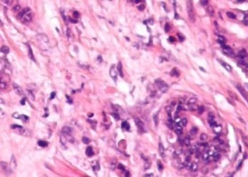
1. Cloacibacterium normanense was significantly enriched in brain tissue samples from those with Alzheimer’s disease compared to controls.
Evidence Rating Level: 2 (Good)
Alzheimer’s disease (AD) is a progressive neurodegenerative disease that is accompanied by memory loss, cognitive impairment, and language difficulties. Several theories have been proposed in explanation of its pathogenesis, but mounting evidence is suggesting a role for neuroinflammatory processes and bacterial infections potentially driving the descent into AD. Specifically, the gut microbiota is known to influence the blood-brain barrier permeability and activate pathways associated with AD progression. Mixed results in previous literature have prompted the current study, which made use of 16S rRNA gene sequencing analysis on thirty brain samples from the frontal cortex and hippocampus, extracted from four AD patients, four controls, and three blank controls. It was found that bacteriotas in brain tissues were differentiated by the brain region rather than by the presence or absence of AD. Taxonomic compositions of brain bacteria revealed no significant phyla differences among the different brain regions. At the genius level, it was found that Cloacibacterium, particularly the Cloacibacterium normanense species, was significantly enriched in the AD group (ps < .01). This species was detected in all four AD patients’ frontal cortexes, and in the hippocampi of two AD patients. This bacteria is found in the human gut and in wastewater. Of the peridontal pathogens in AD brain tissues, only P. gingivalis was detected, but not at significantly higher rates in AD patients compared to controls (p = .888). This study encountered some significant challenges in the amplification of bacterial DNA, and there was more trouble in obtaining valid reads in the hypothalamic and frontal cortex samples. As a result, future studies should focus on improved validation and experimental measures, and explore further the potential role of Cloacibacterium in the brain.
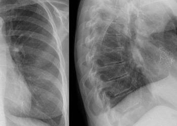
1. In a retrospective cohort analysis, patients with severe SARS-CoV-2 infection were more likely to experience additional symptoms (e.g., fatigue, shortness of breath) in the 31-150 days following a positive test.
2. It was also found that more severe SARS-CoV-2 infections were linked to increased incidence of diabetes, hematologic disorders, and respiratory diseases in the months following a positive test.
Evidence Rating Level: 2 (Good)
The SARS-CoV-2 pandemic has caused millions of infections and deaths worldwide since the first recorded cases. However, there is also a relatively large proportion (between 10 to 50%) of individuals who develop new and persistent symptoms and conditions after infection with SARS-CoV-2 (often referred to as post-acute sequelae of SARS-CoV-2 infection [PASC] or long-COVID). However, these symptoms are heterogenous in nature and have not been appropriately studied in children and non-hospitalized adults. The current study made use of electronic health records data to examine whether select symptoms and conditions could be isolated in relation to acute infection of SARS-CoV-2. Two cohorts were stratified based on age at time of infection (a youth cohort, 0-19 years old, and an adult cohort, ≥ 20 years old). Participants included over 3 million adults (316,249 with a positive viral test and 2,775,331 with only negative viral tests) and over 675,000 youth (62,131 with a positive viral test and 613,512 with only negative tests). Patients who were hospitalized with a positive viral test had higher prevalences of all symptom outcomes 31-150 days after a SARS-CoV-2 test. Fifty-three percent of hospitalized adults with positive tests had at least one symptom compared to 44% of hospitalized adults with negative viral tests. Hospitalized adults with a positive test were at higher risk of being diagnosed with at least one symptom (adjusted odds ratio [aOR], 1.17 [95% CI, 1.11–1.23]), three or more symptoms (aOR, 1.16[95% CI, 1.08 – 1.26]), fatigue (aOR, 1.12 [95% CI, 1.05 – 1.18]), or shortness of breath (aOR, 1.50[95% CI, 1.38–1.63]) 31 to 150 days after SARS-CoV-2 testing. Children who were hospitalized with a positive test had increased odds of being diagnosed with at least one symptom (aOR, 1.18[95% CI, 1.08–1.28]) or shortness of breath (aOR, 1.40 [95% CI, 1.15–1.70]) 31–150 days later. Among those who were not hospitalized, those with positive tests were at higher risk of being diagnosed with fatigue or shortness of breath (aORfatigue, 1.11[95% CI, 1.05–1.16]; aORSOB, 1.22[95% CI, 1.15–1.29]) in the 31-150 days post-positive test. With respect to the development of new conditions, if adults were hospitalized with a positive test, there was an observed increase in incidence of type 1 or 2 diabetes (adjusted hazard ratio [aHR], 1.25[95% CI, 1.17–1.33]), hematologic disorders (aHR, 1.19[95% CI, 1.11–1.28]), and respiratory diseases (aHR, 1.44[95% CI, 1.30–1.60]). They were not significantly more likely than their negative test counterparts to develop mental health conditions, major adverse cardiovascular events, or neurological disorders. If non-hospitalized with a positive test, adults were more likely to develop new hematologic disorders (aHR, 1.12[95% CI, 1.02–1.23]) compared to those who tested negative and did not go to hospital. The generalizability of this study is higher than previous studies due to its inclusion of adults, children, and both hospitalized and non-hospitalized individuals. The findings demonstrate that patients with more severe COVID infections were more likely to develop PASC and long-COVID symptoms. They were also more likely to develop chronic diseases in the months following infection. Future studies should validate these results and further study those who were not hospitalized for long-term sequelae.
Image: PD
©2024 2 Minute Medicine, Inc. All rights reserved. No works may be reproduced without expressed written consent from 2 Minute Medicine, Inc. Inquire about licensing here. No article should be construed as medical advice and is not intended as such by the authors or by 2 Minute Medicine, Inc
![2 Minute Medicine: Pharma Roundup: Price Hikes, Breakthrough Approvals, Legal Showdowns, Biotech Expansion, and Europe’s Pricing Debate [May 12nd, 2025]](https://www.2minutemedicine.com/wp-content/uploads/2025/05/ChatGPT-Image-May-12-2025-at-10_22_23-AM-350x250.png)


