
2 Minute Medicine Rewind March 11, 2024
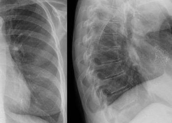
Early vs Late Fixation of Extremity Fractures Among Adults With Traumatic Brain Injury
1. In a cohort of patients with traumatic brain injury, early fixation of extremity fractures within 24 hours of injury was not associated with greater rates of unfavourable functional outcomes at 6 months.
Evidence Rating: 2 (Good)
While extremity fractures are commonly seen in conjunction with traumatic brain injury (TBI), the ideal timing of fixation and definitive treatment of these fractures remain controversial. On one hand, there are those who argue in favour of earlier fixation to promote a shorter recovery course and decreased complication rates. On the other hand however, there are those that argue that the risk of intraoperative complications as well as postoperative stress may further worsen neurologic outcomes. In this multicenter, longitudinal prospective cohort study, data from the Collaborative European NeuroTrauma Effectiveness Research in Traumatic Brain Injury (CENTER-TBI) study was used to further characterize this association. Patients aged 16 or older who underwent internal extremity fixation either within 24 hours or later were propensity score matched to assess differences in unfavourable outcomes at 6 months follow-up. The Glasgow Outcome Scale-Extended (GOSE) score was used as the primary outcome to determine long-term functional outcomes, and secondary outcomes included other measures such as mortality, cardiovascular complications, infections, and other adverse outcomes. Amongst the 253 patients included in the study, which included both mild (N=122) and moderate to severe (N=120) TBI, 74 (29.2%) underwent early internal extremity fixation. It was found that at 6 months follow-up, the rates of both primary and secondary outcomes were not statistically significant between the early fixation for TBI of any severity (odds ratio [OR], 1.12 [95% CI, 0.51-1.99]; P = .77), nor when separating mild from moderate to severe TBI. Overall, although the sample size was small, study findings suggest that it may be safe to provide early intervention for extremity fractures in those with TBI, although further research will be required.
1. In a large prospective cohort, consumption of higher-quality carbohydrates was associated with a decreased risk of colorectal cancer compared to lower quality carbohydrate diets.
2. Lower carbohydrate quantity consumption was not associated with significant correlations in colorectal cancer risk or outcomes.
Evidence Rating: 1 (Excellent)
Colorectal cancer (CRC) confers a significant healthcare burden worldwide, and it has been well documented that individual CRC risk can be mediated through diet. In prior literature, both the quantity and quality of carbohydrate consumption has been postulated as potential mediators of CRC risk and outcomes, although this has not been well characterized. To further characterize this link, patients from the Prostate, Lung, Colorectal, and Ovarian (PLCO) Cancer Screening Trial, a large randomized clinical trial between 1993 and 2001, were followed over a mean follow-up of 8.8 years. They had both the quality and quantity of their carbohydrate consumption tracked, as measured by the carbohydrate quality index (CQI), and low-carbohydrate diet score (LCDs), respectively. CQI was calculated via a validated tool that assessed factors such as glycemic index, whole grain to total grain ratio, solid carbohydrate to total carbohydrate ratio, and dietary fiber. It was found that individuals in the highest quartiles of CQI had significant lower CRC incidence (HR 0.80 95% CI 0.67–0.96) and mortality (Q4 vs Q1: HR 0.61, 95% CI 0.44–0.86) when compared to those in the lowest quartiles, specifically for the distal colon and rectum. Interestingly, no associations were found between LCDs and CRC incidence or mortality in any CRC types. Study findings suggest that quality of carbohydrate intake, rather than quantity, has the effect on CRC incidence as well as mortality risk. With further research, practitioners and dietitians alike may utilize study findings to help counsel patients on their dietary choices to further mitigate CRC risk.
Incidence of nonkeratinocyte skin cancer after breast cancer radiation therapy
1. Amongst a cohort of breast cancer survivors, those who were treated with radiation had a higher risk of developing nonkeratinocyte skin cancer.
Evidence Rating Level: 2 (Good)
Cancer remains the second leading cause of death in the United States (US), even though incidence rates have declined. In the US, there are over 4 million breast cancer survivors that deal with mental and physical health challenges after the end of their treatment. Of the 875 880 patients with a new diagnosis of breast cancer, 51.6% were 60 years of age or older and 50.3% underwent radiation therapy. Further, Of the participants, 11.2% identified as Hispanic, 10.1% identified as non-Hispanic Black, and 69.5% identified as non-Hispanic White. Standardized incidence ratios (SIRs) for subsequent nonkeratinocyte skin cancer development based on treatment type, skin cancer location, and skin cancer subtype, were the primary outcomes of the study. Between 2000 and 2019, a total of 3839 patients who were finished with treatment developed nonkeratinocyte cancer, which includes melanoma (3419 [89.1%]), Merkel cell carcinoma (121 [3.2%]), hemangiosarcoma (104 [2.7]) and 32 others (195 [5.1%]). When compared to the general population, the risk of getting diagnosed with nonkeratinocyte skin cancer was 57% higher (SIR, 1.57 [95% CI, 1.45-1.7]) in those who received breast cancer treatment. When compared to the general population, the incidence of melanoma increased by 1.37-fold (SIR, 1.37 [95% CI, 1.25-1.49]) and the incidence of hemangiosarcoma increased by 27.11-fold (SIR, 27.11 [95% CI, 21.6-33.61]) when localized to the skin of the breast or trunk. Different cancer treatments had differing risks of nonkeratinocyte skin cancer, with radiation having a much greater risk than chemotherapy and surgery. The increased risk of developing nonkeratinocyte skin cancer in breast cancer survivors was statistically significant, with an even greater risk in those that received radiation as their treatment. Consistent with previous data, the findings of the study showed the role of radiation as a risk for secondary malignant neoplasms. Overall, breast cancer survivors had a greater risk of developing melanoma and hemangiosarcoma after treatment with radiation. Although nonkeratinocyte cancers are rare, it is important for physicians to understand their elevated risk of occurrence in breast cancer survivors previously treated with radiation.
1. Pregnant individuals with obesity according to their body mass index (BMI) had an increased risk for stillbirth when compared to pregnant individuals with a normal BMI
Evidence Rating Level: 2 (Good)
While obesity has been shown to be a risk factor for stillbirths, there may be conflating factors contributing to these findings. Among pregnant people with obesity, advancing gestational age was associated with increased hazard ratios (HRs). A retrospective cohort study was undertaken to examine the effects of additional factors compared to obesity in pregnant people alone as a risk factor for stillbirths. The Better Outcomes Registry and Network (BORN) provided data for antenatal, intrapartum, postpartum, and neonatal outcomes for births that occurred after 20 weeks’ gestation in Ontario, Canada. To understand the relationship between prepregnancy body mass index (BMI) class and stillbirth, multivariable Cox proportional hazard regression analysis was used as it was able to control for possible confounding variables. Logistic regression was also used to examine odds ratios (ORs) and 95% confidence intervals (CIs) for stillbirths. For the analysis, obstetrical complications were treated independently from maternal characteristics. Mediator analyses were used to examine the effects of BMI on stillbirths. The results were modeled to examine whether maternal obesity was the sole risk factor for stillbirth. The data pool for this study included 681 178 births that occurred between 2012 and 2018, of which 1956 were stillbirths. There was an association between Class I obesity and an increased occurrence of stillbirth (adjusted hazard ratio [HR] 1.55, 95% confidence interval [CI] 1.35–1.78). However, class III obesity was associated with an even higher incidence (adjusted HR 1.80, 95% CI 1.44–2.24), and class II with the highest incidence of stillbirths (adjusted HR 2.17, 95% CI 1.83–2.57). Compared to those with a normal BMI, those with obesity as their only risk factor had an increase chance of stillbirth past 37 weeks’ gestation as found by plotting point estimated for odds ratios. Pregnant people experiencing obesity, especially the highest classes, have an increased risk of stillbirths when compared to pregnant people with a normal BMI. This risk further increases with advancing gestational age. Overall, there was a higher risk of stillbirths associated with maternal obesity even after adjusting for prevalent risk factors in this community.
1. In a large prospective multicentre diagnostic accuracy study, magnetically guided capsule endoscopy with a detachable string (ds-MCE) demonstrated high accuracy and safety in the detection and grading of esophagogastric varcies in patients with cirrhosis.
Evidence Rating: 1 (Excellent)
Esophagogastric varices are highly prevalent in patients with cirrhosis and are a major cause of mortality and morbidity. While capsule endoscopy has been proposed as a standard to esophagoduodenoscopy (EGD) in the past, prior studies have demonstrated an inferior accuracy rating compared to EGD, owing to a poor visualization of the esophagus and stomach due to rapid transit times. To rectify this, researchers developed a ds-MCE and enrolled patients in a multicentre prospective study to assess its diagnostic accuracy. 607 adult patients with cirrhosis were recruited, and had ds-MCE followed by standard EGD. The sensitivity and specificity of ds-MCE for the detection of esophagogastric varices were then compared to EGD via diagnostic accuracy testing. When compared to EGD as the reference standard, ds-MCE demonstrated a sensitivity of 97.5% and specificity of 97.8%, without occurrence of any serious adverse events. This compares to a historical sensitivity of 74% in one prior multicentre study. Overall, the modification of standard capsule endoscopy with a ds-MCE appeared to be an effective, and safe alternative to EGD in the detection of esophagogastric varices in patients with cirrhosis. In patients that would be poor candidates for sedation, or who would otherwise not tolerate EGD, ds-MCE may present itself as an effective alternative diagnostic tool.
Image: PD
©2024 2 Minute Medicine, Inc. All rights reserved. No works may be reproduced without expressed written consent from 2 Minute Medicine, Inc. Inquire about licensing here. No article should be construed as medical advice and is not intended as such by the authors or by 2 Minute Medicine, Inc.


